
Cell - David Orloff
@CellImageLibrar
The Cell is a public and easily accessible resource database of images, videos, and animations of #cells and #microscopy. Donate a tweet - http://bit.ly/yIYVNk
You might like


📷 Image of the Week – June 22, 2020 CIL:11397 - cellimagelibrary.org/images/11397 Description: Figure 218 from Chapter 7 (Mitochondria) of ‘The Cell, 2nd Ed.’ by Don W. Fawcett M.D. The typical structural organization of mitochondria... tmblr.co/ZsZBfwYWUydLie…
cellimagelibrary.tumblr.com
cellimagelibrary
Image of the Week – June 22, 2020 CIL:11397 - http://cellimagelibrary.org/images/11397 Description: Figure 218 from Chapter 7 (Mitochondria) of ‘The Cell, 2nd Ed.’ by Don W. Fawcett M.D. The typica...
📷 Cheers! Image of the Week – June 15, 2020 CIL:41635 - cellimagelibrary.org/images/41635 Description: Fluorescent image of the sporangium, an enclosure in which spores are formed, of the slime mold Craterium minutum. Honorable... tmblr.co/ZsZBfwYUEVAP0i…
cellimagelibrary.tumblr.com
cellimagelibrary
Cheers! Image of the Week – June 15, 2020 CIL:41635 - http://www.cellimagelibrary.org/images/41635 Description: Fluorescent image of the sporangium, an enclosure in which spores are formed, of the...
📹 Image of the Week – June 8, 2020 CIL:12375 - cellimagelibrary.org/images/12375 Description: Movie showing the dynamics of kinetchore microtubules during meiosis II in primary spermatocytes of the crane-fly Nephrotoma suturalis that... tmblr.co/ZsZBfwYR_UQoee…
📹 Image of the Week – June 1, 2020 CIL:50519 - cellimagelibrary.org/images/50519 Description: We examined the structural organization of cytoskeletal components and membrane systems in neurons of well preserved biopsy material from... tmblr.co/ZsZBfwYPk5hgGq…
You’ve Got Some Nerve! Image of the Week – 2/17/20 cellimagelibrary.org/images/41833 Timothy Mosca and 2010 Olympus BioScapes Digital Imaging Competition® CC - by-nc-nd

📷 You’ve Got Some Nerve! Image of the Week – February 17, 2020 CIL:41833 - flagella.crbs.ucsd.edu/images/41833 Description: Confocal micrograph showing the complex connectivity at the neuromuscular junction of Drosophila (fruit fly).... tmblr.co/ZsZBfw2nlYKLw
📷 You Dirty Rat! Image of the Week – February 10, 2020 CIL:48107 - cellimagelibrary.org/images/48107 Description: Differentiated rat neural stem cells stained for β III-Tubulin (in blue) to reveal neurons and GFAP (in red) to reveal... tmblr.co/ZsZBfw2neIif5
📷 Dinosaur! Image of the Week – February 3, 2020 CIL:41919 - cellimagelibrary.org/images/41919 Description: Polarized light micrograph of Triceratops dinosaur parietal (skull) bone. Honorable Mention, 2009 Olympus BioScapes Digital... tmblr.co/ZsZBfw2nWjpAH

📷 Image of the Week – January 27, 2020 CIL:45553 - cellimagelibrary.org/images/45553 Description: Imaging an HIV infected dendritic cell reveals numerous viral particles at the tips of filopodia. Actin-based membrane extensions from the... tmblr.co/ZsZBfw2nOv1-Z

📷 Coronavirus! - Image of the Week – January 20, 2020 CCDB:6022 - cellimagelibrary.org/images/CCDB_60… Project name: Membrane Modifications Induced by Corona Virus Description: Electron tomography of African Green Monkey... tmblr.co/ZsZBfw2nH5465
cellimagelibrary.tumblr.com
cellimagelibrary
Coronavirus! - Image of the Week – January 20, 2020 CCDB:6022 - http://www.cellimagelibrary.org/images/CCDB_6022 Project name: Membrane Modifications Induced by Corona Virus Description: Electron...
📷 Image of the Week - January 6, 2020 CIL:34603 - cellimagelibrary.org/images/34603 Description: A cross section of cilium through the cilium-basal body complex extending from the cilium to the transition zone between the cilium and... tmblr.co/ZsZBfw2n14Sxs

📷 Image of the Week - December 30, 2019 CIL:41488 - cellimagelibrary.org/images/41488 Description: Colorized scanning electron micrograph of pollen (blue) in anther (green) held by the filament (pink ) from Acacia dealbata grown in... tmblr.co/ZsZBfw2murQ7H
cellimagelibrary.tumblr.com
cellimagelibrary
Image of the Week - December 30, 2019 CIL:41488 - http://www.cellimagelibrary.org/images/41488 Description: Colorized scanning electron micrograph of pollen (blue) in anther (green) held by the...
📷 Image of the Week – December 16, 2019 CIL:11397 - cellimagelibrary.org/images/11397 Description: Figure 218 from Chapter 7 (Mitochondria) of ‘The Cell, 2nd Ed.’ by Don W. Fawcett M.D. The typical structural organization of... tmblr.co/ZsZBfw2mfiMVQ

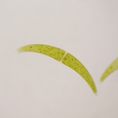
📷 Find this and other great images in the Technology Networks new The Spectacular World of Aquatic Organisms Flipbook. Image of the Week - November 25, 2019 CIL:41017 - cellimagelibrary.org/images/41017 Description: Laser scanning... tmblr.co/ZsZBfw2mGswUJ

📷 Image of the Week – November 18, 2019 CIL:41311 - cellimagelibrary.org/images/41311 Description: Scanning electron microscope image of Penta lanceolata stigma (receptive surface for pollen). Authors: Louisa Howard and Charles Daghlian... tmblr.co/ZsZBfw2m8URiD
cellimagelibrary.tumblr.com
cellimagelibrary
Image of the Week – November 18, 2019 CIL:41311 - http://cellimagelibrary.org/images/41311 Description: Scanning electron microscope image of Penta lanceolata stigma (receptive surface for...
📷 Image of the Week – November 11, 2019 CIL:50647 - cellimagelibrary.org/images/50647 Description: Human umbilical vein endothelial cells stained initially for nuclei with DAPI (blue) and for vascular endothelial cadherin (red).... tmblr.co/ZsZBfw2l-MIkP

📷 Spooky Eyes - Happy Halloween - Image of the Week - October 28, 2019 CIL:39022 - cellimagelibrary.org/images/39022 Description: Confocal micrograph showing the connections of the visual system in a four-day-old zebrafish embryo.... tmblr.co/ZsZBfw2liA7og

📷 Image of the Week – October 21, 2019 CIL:48108 - cellimagelibrary.org/images/48108 Description: Differentiated rat neural stem cells stained for β III-Tubulin (in green) to reveal neurons, GFAP (in red) to reveal glial cells, and DAPI... tmblr.co/ZsZBfw2lXIIGQ

📷 Image of the Week - September 16, 2019 CIL:38814 - cellimagelibrary.org/images/38814 Description: A scanning electron color-enhanced image showing a clump of prostate cancer cells. Authors: Annie Cavanagh Licensing:... tmblr.co/ZsZBfw2ksV4zV


United States Trends
- 1. Kevin James 1,324 posts
- 2. Bubba 25K posts
- 3. Bill Clinton 107K posts
- 4. #CashAppGreen 1,260 posts
- 5. #BravoCon 3,540 posts
- 6. Hayley 18K posts
- 7. Hunter Biden 17.8K posts
- 8. Wale 45.3K posts
- 9. Metroid 9,662 posts
- 10. Rondo 2,598 posts
- 11. John Beam 4,318 posts
- 12. Crooks 75K posts
- 13. Vatican 13.7K posts
- 14. Jaylon Johnson 2,218 posts
- 15. RIP Coach Beam N/A
- 16. #FanCashDropPromotion 3,792 posts
- 17. #FursuitFriday 14.4K posts
- 18. Last Chance U 2,256 posts
- 19. Paul Blart N/A
- 20. Bondi 99.7K posts
You might like
-
 EMBO
EMBO
@EMBO -
 American Society for Cell Biology
@ASCBiology -
 HHMI
HHMI
@HHMINEWS -
 Allen Institute
@AllenInstitute -
 Microscopy Society
@MicroscopySoc -
 nationalpostdoc
@nationalpostdoc -
 BioImagingUK
@BioImagingUK -
 Biomedical Picture of the Day BPoD
@BPoD_s -
 Paul Kenny, PhD
@PaulKennyPhD -
 Christophe Leterrier
@christlet -
 Leica Microsystems
@LeicaMicro -
 Professor Brian D Smith, Academic, Author, Advisor
@ProfBrianDSmith -
 Helen Rizos
@HelenRizos -
 Vanderbilt Center for Structural Biology
@VanderbiltCSB -
 Shengqian Ma@UNT
@shengqianma
Something went wrong.
Something went wrong.