
You might like


Continuing our series on finger injuries... Next moving on to the PIP JOINT... NORMAL ANATOMY :- The PIP joint comprises of the following stabilizing structures: • Collateral ligament complex- collateral ligament proper and an accessory collateral ligament (similar to MCP…


PECTORALIS MAJOR TENDON: 2 layers: •ANTERIOR: Clavicular & superior Sternal segments •POSTERIOR: remaining Sternal segments Slides from my last lecture at @RadiologiaChile congress


Midline RA/AL pubic plate injury. Mri versus US/illustration correlation. #mskrad

Rectus abdominis/adductor longus common aponeurotic injury. Correlation MRI versus US/illustration. #mskrad

We try to call on MRI but, we can’t compete with dynamic ultrasound like this! Fantastic imaging.

Enteric duplication cyst with bowel mucosa sign.

Absolutely wonderful figure showing the spectrum of radiologic imaging techniques for evaluating injury to the shoulder girdle👍👏👏 From :Orthopedic Imaging A Practical Approach 6th Edi, Adam Greenspan et al, Lippincott Williams & Wilkins/ Wolters Kluw Health

♦️os trigonum has rounded with corticated edges. VS ♦️Stieda process Post process of talus has (medial tubercle) & (lateral tubercle= Stieda’s process). VS ♦️Fx of posterolateral process of talus (Shepherd’s Fx) canbe mistaken as os trigonum Image: pin.it/6Zt7MAh3r


When your patient starts to crump—remember CRUMPLED! Cortical restricted diffusion on MRI is often seen in neurologic decline. Do you crumple under the pressure of trying to remember all the causes of cortical restricted diffusion? Never fear! Here’s an easy mnemonic to help…

1/Wish that your knowledge of autoimmune encephalitis was automatic? Do you feel in limbo when it comes to the causes of limbic encephalitis? Do you know the patterns of autoimmune encephalitis? Here’s a thread with some hints to help you figure it all out!

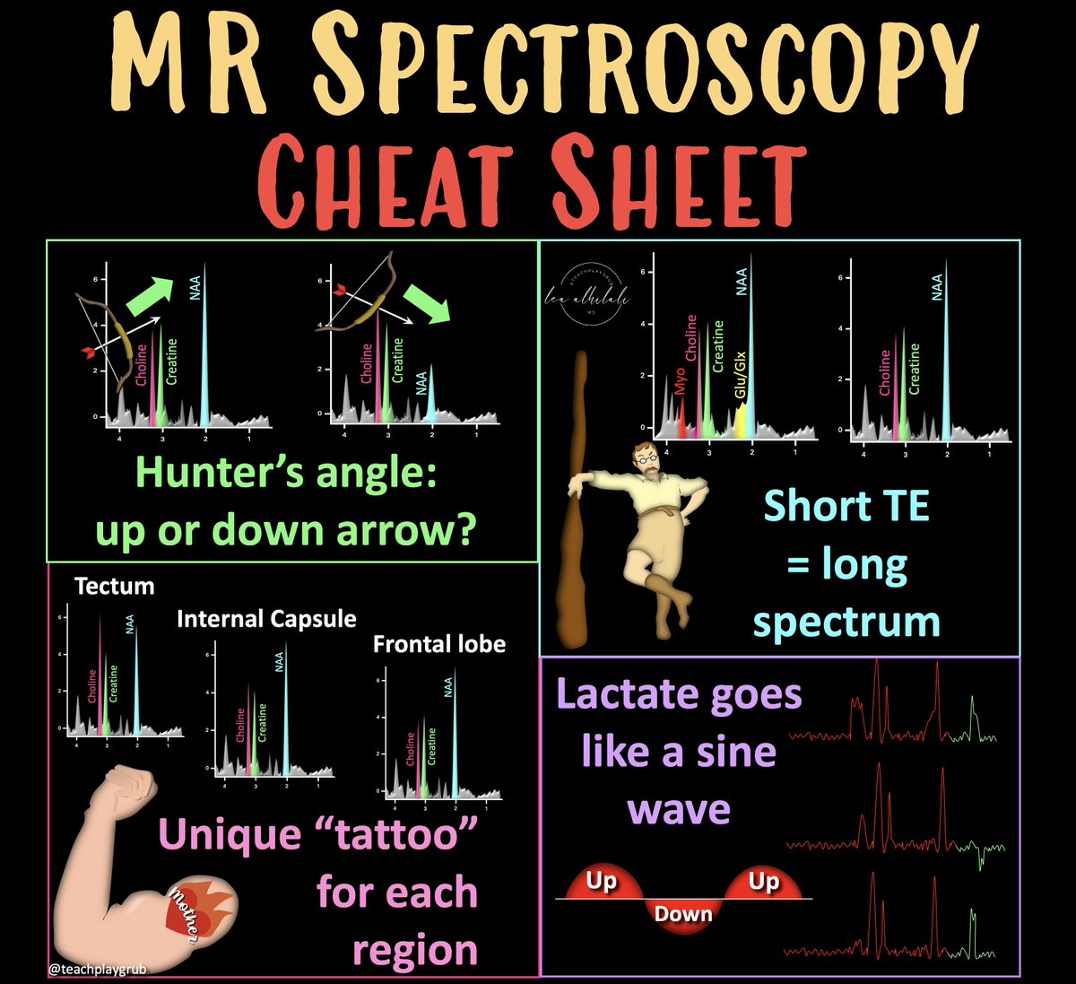
1/Tired of always speculating about MR spectroscopy? If you've ever looked at an MR spectroscopy & thought: "I have no idea what I’m looking at!"--then this cheat sheet is for you! Here's a thread on the 4 basic rules you need to understand the spectrum of basic spectroscopy!
Now you know the three planes & three types of compression to look for when you are looking at the fifth nerve. Hopefully, now the imaging of trigeminal neuralgia won't cause you any more pain!

2/The most important thing to remember is that the nerve is 3D so you have to look at it in all three planes. So what is the normal and abnormal appearance of the trigeminal nerve in each plane?

Are you pretty bad about knowing when to give gad? Do you know which gadolinium contrast is safe to give? Know what to do w/gadolinium in patients w/kidney disease? Here’s the gadolinium cheat sheet you NEED from @theAJNR SCANtastic! Read on... ajnr.org/content/45/8/1… ➡️RISK…

12/For moderate stenosis, you lose the space on all 4 sides, but nerve itself is not compressed or deformed Like a sleek outfit, it shows your curves, but doesn’t deform them. It’s not a comfy outfit, per se, and I wouldn’t eat a lot while wearing it, but it’s not too small

10/For foraminal narrowing, the nerve inside the foramen has fat around it on four sides that gets attenuated as the space gets tighter. How many sides have attenuated fat determines how severe the stenosis is.

11/Mild stenosis is where you have loss of the fat on 2 sides. So it is still comfy clothing bc the fat is preserved on the other two sides, so you have lots of space.

Worried that hydrocephalus is hiding in plain sight? Normal pressure hydrocephalus (NPH) can mimic volume loss How can you tell the difference? This month’s @theAJNR SCANtastic gives you want you need to know ajnr.org/content/45/10/… Many findings can be seen with NPH—you may…

To call it or not to call it? That is the question! Feeling wacky & wobbly when it comes to normal pressure hydrocephalus? Don’t want to overcall it, but don’t want to miss it either! Check out the latest in NPH w/this month’s @theAJNR SCANtastic! ajnr.org/content/45/10/……


United States Trends
- 1. Lakers 50.1K posts
- 2. #AEWDynamite 45.5K posts
- 3. Epstein 1.5M posts
- 4. Jokic 16.1K posts
- 5. Shai 14.8K posts
- 6. #AEWBloodAndGuts 5,649 posts
- 7. #Survivor49 3,664 posts
- 8. Darby 5,414 posts
- 9. Kyle O'Reilly 1,829 posts
- 10. Steph 25.6K posts
- 11. Thunder 41.6K posts
- 12. Rory 7,318 posts
- 13. Moxley 2,883 posts
- 14. Kobe Sanders N/A
- 15. Spencer Knight N/A
- 16. Hobbs 28.6K posts
- 17. Caruso 4,023 posts
- 18. #SistasOnBET 2,255 posts
- 19. Warriors 49.5K posts
- 20. Blood & Guts 25K posts
You might like

Something went wrong.
Something went wrong.