#cellsegmentation search results
Once we have a stitched image, how do we discover single cell phenotypes that interest us? ➡️ SPARCSpy 🔵 SPARCSpy performs precise cell segmentation, generating single-cell image datasets for all cells in a sample #cellsegmentation #singlecellimagedataset


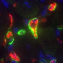
🎯 Precise #CellSegmentation is challenging. @vizgen_inc uses cell boundary stains and robust algorithms for accuracy. See these #DataImages of FFPE mouse breast tumor by researchers at @NeuroAlc, showcasing cell boundary staining and segmentation revealing natural cell shapes.
#Baysor #CellSegmentation for 2D 3D image-based #SpatialTranscriptomics Based on Transcript composition #MarkovRandomField -/+Stain image #PriorSegmentationConfidence Work with #MERFISH #smFISH #STARmap #InSituSequencing ⏫Detection of #EndothelialCell #MuralCell (Fig 5/6)…

Customize your #CellSegmentation with the new #Cellpose2 Plugin for the #Vizgen Post-Processing Tool! Visit our website to learn how you can improve segmentation results for challenging tissues with the plugin for #VPT: hubs.ly/Q02k2S000 #MERSCOPE #MERFISH #Spatialomics

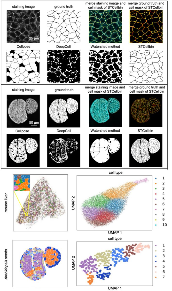
✨Our latest blog tests 8 popular community-developed tools using Stereo-seq data. Discover which algorithms excel across diverse staining types and explore segmentation-free methods for special cases! Dive in: bit.ly/3Vnn2D7 #CellSegmentation #Spatialbiology #STOmics

🍬Treat yourself this Halloween with the new #Vizgen Post-Processing Tool for Cell Segmentation! #VPT uses #CellSegmentation methods to draw cell boundaries & generates #SingleCell output by combining the results with #MERFISH data. Learn more: hubs.ly/Q026kTq70 #MERSCOPE
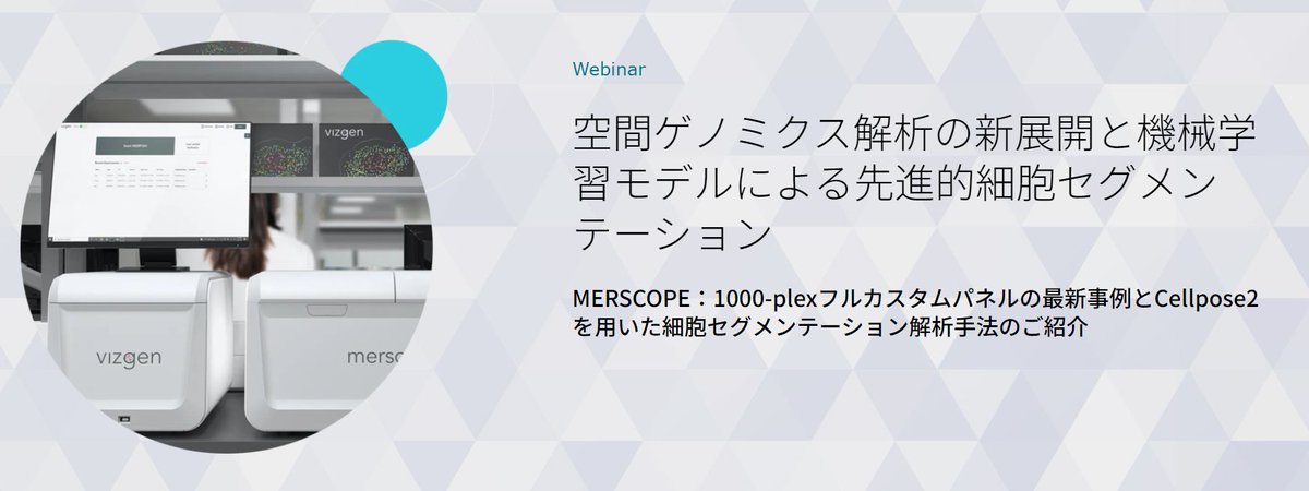
💻Webinar Alert for #SpatialTranscriptomics Customers in Japan! Developments in Spatial Genomics Analysis and Advanced Cell Segmentation 📅 Wednesday, April 24, 2024 ⏰15:00 - 15:45 JST 🎙️Wenbin Gu, Primetech Co. 🔗 hubs.ly/Q02sQSrr0 #CellSegmentation #MERSCOPE

Get a closer look at the superior cell segmentation and transcript information you can obtain from CosMx SMI data during our upcoming Spatial Informatics 101 Webinar on July 11. Register to attend: bit.ly/3pxrlQ4 #SpatialInformatics #CellSegmentation

In October '21, researchers from @Harvard, @BostonChildrens, & @uni_copenhagen published a paper in @NatureBiotech describing a new method for #CellSegmentation called #Baysor. Read & learn how they evaluated it with #MERFISH! hubs.ly/Q013pM8Q0 #ThrowbackThursday

What to do when #cellsegmentation does not properly work and creates an artificial #cellmask? One can always make something else out of it😊


We are very impressed with the ease of creation and quality of this open source @DeepCell_Inc #CellSegmentation mask made in Steinbock in the @BodenmillerLab. Our #ImagingMassCytometry data analysis is much improved!


Working on cell segmentation in colon tissue @MikeKattahMD @iuliarusu9 @JaredBain909 #cellsegmentation #spatialbiology #colon #colorful #ucsf

STCellbin Use Nuclei+Cell Membrane/Wall stains to improve #CellSegmentation & single-cell-resolved #SpatialTranscriptomics Tailored for #StereoSeq, but likely implementable for other ST methods Susanne Brix & Xun Xu labs @GigaByteJournal 2024 gigabytejournal.com/articles/110
Welcome to the 6th day of 12 Days of NanoString! Interested in leaping to single-cell spatial in these applications? nanostring.com/events/spatial… #SpatialData #CellSegmentation #SpatialTranscriptomics #SingleCellImaging


DON'T MISS Dr. McArdle's @pmlsummit talk this Thurs 9/14 featuring Orion data utilizing @QuPath for #imagevisualization, #cellsegmentation via #deeplearningmodels, #cellphenotyping, and #spatialanalysis. For free registration: rarecyte.com/contact-us/ #spatialbiology

On the final day of 12 Days of NanoString unwrap all of our spatial technologies. Watch the 5th Annual Spatial Genomics Summit highlighted diverse spatial applications. bit.ly/3GEAXgQ #SingleCellImaging #SpatialMultiomics #CellSegmentation #SpatialBiology


RAMCES, a computational method by @monica_dayao et al., improves #cellsegmentation in imaging-based #spatialproteomics by facilitating the selection of optimal membrane protein markers @CMUPittCompBio @CMUCompBio go.nature.com/37TVoZg


Ko Sugawara shows how training with sparse annotations can produce practical models for cell image segmentation. Strategic selection of annotated cells boosts performance! @ELEPHANT_track @NEUBIAS_ Symposium 🧪🔍 #DeepLearning #BioimageAnalysis #CellSegmentation

The product of the study ""Real-Time Three-Dimensional #cellsegmentation in Large-Scale Microscopy Data of Developing #embryos"" (available at sciencedirect.com/science/articl…) can be observed throught #MorphoNet. #imagevisualization #CellBiology 🔬
Dr. McArdle's @pmlsummit talk on 9/14 will feature Orion data being used with @QuPath for #imagevisualization, #cellsegmentation via #deeplearningmodels, #cellphenotyping, and #spatialanalysis. For free conference registration: rarecyte.com/contact-us/ #spatialbiology

✨Our latest blog tests 8 popular community-developed tools using Stereo-seq data. Discover which algorithms excel across diverse staining types and explore segmentation-free methods for special cases! Dive in: bit.ly/3Vnn2D7 #CellSegmentation #Spatialbiology #STOmics

🔬 Reducing annotation costs in #cellsegmentation📉 Study shows how a small, annotated dataset can train a model to upgrade low-quality annotations, improving segmentation accuracy and efficiency. 💡mdpi.com/2313-433X/10/7… #MDPIJimaging #DeepLearning #ComputerVision


🔬 Reducing annotation costs in #cellsegmentation📉 Study shows how a small, annotated dataset can train a model to upgrade low-quality annotations, improving segmentation accuracy and efficiency. 💡mdpi.com/2313-433X/10/7… #MDPIJimaging #DeepLearning #ComputerVision


#cellsegmentation and classification are critical tasks in spatial #omics data analysis. Unlike the typical two-stage approach of segmentation followed by classification, #CelloType implements multitask learning strategy that integrates the two, enhancing the performance of both.
🎯 Precise #CellSegmentation is challenging. @vizgen_inc uses cell boundary stains and robust algorithms for accuracy. See these #DataImages of FFPE mouse breast tumor by researchers at @NeuroAlc, showcasing cell boundary staining and segmentation revealing natural cell shapes.
🎯 Precise #CellSegmentation is challenging. @vizgen_inc uses cell boundary stains and robust algorithms for accuracy. See these #DataImages of FFPE mouse breast tumor by researchers at @NeuroAlc, showcasing cell boundary staining and segmentation revealing natural cell shapes.
💻Webinar Alert for #SpatialTranscriptomics Customers in Japan! Developments in Spatial Genomics Analysis and Advanced Cell Segmentation 📅 Wednesday, April 24, 2024 ⏰15:00 - 15:45 JST 🎙️Wenbin Gu, Primetech Co. 🔗 hubs.ly/Q02tBwP_0 #CellSegmentation #MERSCOPE

💻Webinar Alert for #SpatialTranscriptomics Customers in Japan! Developments in Spatial Genomics Analysis and Advanced Cell Segmentation 📅 Wednesday, April 24, 2024 ⏰15:00 - 15:45 JST 🎙️Wenbin Gu, Primetech Co. 🔗 hubs.ly/Q02sQSrr0 #CellSegmentation #MERSCOPE
STCellbin Use Nuclei+Cell Membrane/Wall stains to improve #CellSegmentation & single-cell-resolved #SpatialTranscriptomics Tailored for #StereoSeq, but likely implementable for other ST methods Susanne Brix & Xun Xu labs @GigaByteJournal 2024 gigabytejournal.com/articles/110
Customize your #CellSegmentation with the new #Cellpose2 Plugin for the #Vizgen Post-Processing Tool! Visit our website to learn how you can improve segmentation results for challenging tissues with the plugin for #VPT: hubs.ly/Q02mN7ZV0 #MERSCOPE #MERFISH #SpatialOmics
Customize your #CellSegmentation with the new #Cellpose2 Plugin for the Vizgen Post-Processing Tool! Visit our website to learn more about #VPT and the Cellpose2 cell segmentation plugin: hubs.ly/Q02m2Xf80 #Vizgen #MERSCOPE #MERFISH #Spatialomics

✨Our latest blog tests 8 popular community-developed tools using Stereo-seq data. Discover which algorithms excel across diverse staining types and explore segmentation-free methods for special cases! Dive in: bit.ly/3Vnn2D7 #CellSegmentation #Spatialbiology #STOmics
In October '21, researchers from @Harvard, @BostonChildrens, & @uni_copenhagen published a paper in @NatureBiotech describing a new method for #CellSegmentation called #Baysor. Read & learn how they evaluated it with #MERFISH! hubs.ly/Q013pM8Q0 #ThrowbackThursday
#Baysor #CellSegmentation for 2D 3D image-based #SpatialTranscriptomics Based on Transcript composition #MarkovRandomField -/+Stain image #PriorSegmentationConfidence Work with #MERFISH #smFISH #STARmap #InSituSequencing ⏫Detection of #EndothelialCell #MuralCell (Fig 5/6)…
STCellbin Use Nuclei+Cell Membrane/Wall stains to improve #CellSegmentation & single-cell-resolved #SpatialTranscriptomics Tailored for #StereoSeq, but likely implementable for other ST methods Susanne Brix & Xun Xu labs @GigaByteJournal 2024 gigabytejournal.com/articles/110
💻Webinar Alert for #SpatialTranscriptomics Customers in Japan! Developments in Spatial Genomics Analysis and Advanced Cell Segmentation 📅 Wednesday, April 24, 2024 ⏰15:00 - 15:45 JST 🎙️Wenbin Gu, Primetech Co. 🔗 hubs.ly/Q02sQSrr0 #CellSegmentation #MERSCOPE
Welcome to the 6th day of 12 Days of NanoString! Interested in leaping to single-cell spatial in these applications? nanostring.com/events/spatial… #SpatialData #CellSegmentation #SpatialTranscriptomics #SingleCellImaging
Get a closer look at the superior cell segmentation and transcript information you can obtain from CosMx SMI data during our upcoming Spatial Informatics 101 Webinar on July 11. Register to attend: bit.ly/3pxrlQ4 #SpatialInformatics #CellSegmentation
Customize your #CellSegmentation with the new #Cellpose2 Plugin for the Vizgen Post-Processing Tool! Visit our website to learn more about #VPT and the Cellpose2 cell segmentation plugin: hubs.ly/Q02m2Xf80 #Vizgen #MERSCOPE #MERFISH #Spatialomics
Working on cell segmentation in colon tissue @MikeKattahMD @iuliarusu9 @JaredBain909 #cellsegmentation #spatialbiology #colon #colorful #ucsf
On the final day of 12 Days of NanoString unwrap all of our spatial technologies. Watch the 5th Annual Spatial Genomics Summit highlighted diverse spatial applications. bit.ly/3GEAXgQ #SingleCellImaging #SpatialMultiomics #CellSegmentation #SpatialBiology
Once we have a stitched image, how do we discover single cell phenotypes that interest us? ➡️ SPARCSpy 🔵 SPARCSpy performs precise cell segmentation, generating single-cell image datasets for all cells in a sample #cellsegmentation #singlecellimagedataset
RAMCES, a computational method by @monica_dayao et al., improves #cellsegmentation in imaging-based #spatialproteomics by facilitating the selection of optimal membrane protein markers @CMUPittCompBio @CMUCompBio go.nature.com/37TVoZg
Dr. McArdle's @pmlsummit talk on 9/14 will feature Orion data being used with @QuPath for #imagevisualization, #cellsegmentation via #deeplearningmodels, #cellphenotyping, and #spatialanalysis. For free conference registration: rarecyte.com/contact-us/ #spatialbiology
DON'T MISS Dr. McArdle's @pmlsummit talk this Thurs 9/14 featuring Orion data utilizing @QuPath for #imagevisualization, #cellsegmentation via #deeplearningmodels, #cellphenotyping, and #spatialanalysis. For free registration: rarecyte.com/contact-us/ #spatialbiology
Ko Sugawara shows how training with sparse annotations can produce practical models for cell image segmentation. Strategic selection of annotated cells boosts performance! @ELEPHANT_track @NEUBIAS_ Symposium 🧪🔍 #DeepLearning #BioimageAnalysis #CellSegmentation
What to do when #cellsegmentation does not properly work and creates an artificial #cellmask? One can always make something else out of it😊
We are very impressed with the ease of creation and quality of this open source @DeepCell_Inc #CellSegmentation mask made in Steinbock in the @BodenmillerLab. Our #ImagingMassCytometry data analysis is much improved!
🔬 Reducing annotation costs in #cellsegmentation📉 Study shows how a small, annotated dataset can train a model to upgrade low-quality annotations, improving segmentation accuracy and efficiency. 💡mdpi.com/2313-433X/10/7… #MDPIJimaging #DeepLearning #ComputerVision

Cell Segmentation is the task of splitting a microscopic image domain into segments, which represent individual instances of cells. For more updates follow: lnkd.in/daSxGfTR Submit your work: [email protected] #cellsegmentation #cellbiology #microscopy

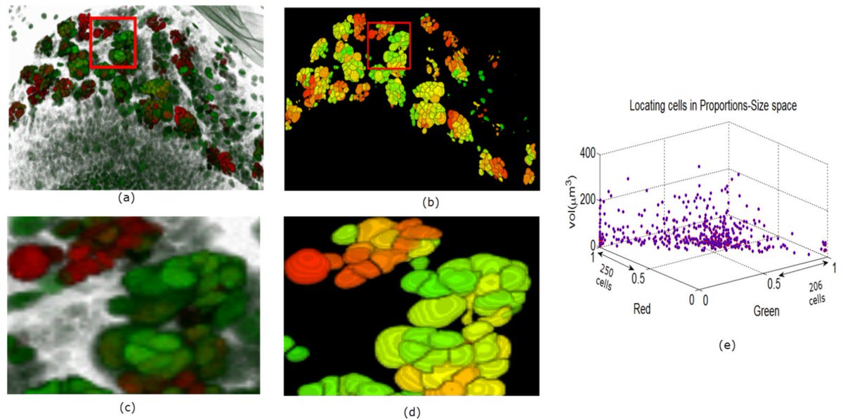
#3D Clumped #CellSegmentation Using Curvature Based Seeded #Watershed. Full text free at: mdpi.com/2313-433X/2/4/… #imaging #openaccess
Something went wrong.
Something went wrong.
United States Trends
- 1. #WorldSeries 139K posts
- 2. #SNME 74.1K posts
- 3. Ohtani 59.6K posts
- 4. Blue Jays 80.6K posts
- 5. Hugh Freeze 2,181 posts
- 6. Auburn 8,516 posts
- 7. Gimenez 14.8K posts
- 8. Bo Bichette 23.2K posts
- 9. Jesse Love 3,127 posts
- 10. Mateer 2,570 posts
- 11. Jordan Marshall 1,300 posts
- 12. Zilisch 4,847 posts
- 13. Max Scherzer 12.8K posts
- 14. Shohei 41.9K posts
- 15. Wrobleski 6,667 posts
- 16. Toronto 50.3K posts
- 17. Purdue 4,274 posts
- 18. CM Punk 25.9K posts
- 19. #UFCVegas110 14.8K posts
- 20. Vlad 8,549 posts