
bittesla.sol (standard and poor)
@BitTesla
Doctor trying not to be a Dollar pleb. Learning finance also saves lives. Trade at GMX dex with no KYC http://gmx.io/?ref=nokyc
You might like


Reminder for self

5.1/ For beginners who cannot cut losses, close your positions and immediately reopen them. Your PNL on that position resets, and this exercise helps to clear biases from loss aversion. Still want to keep them on?

Get ready, Dr. Usha Nagaraj is about to teach you everything you need to know about sellar, suprasellar, pineal, and intraventricular pediatric tumors. #ASPNR26


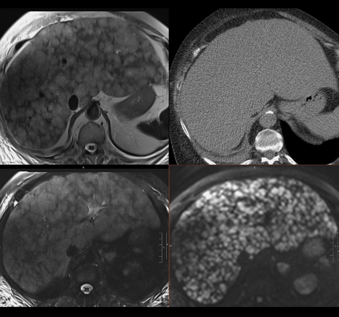
Good reminder that anything can be hiding in the liver on that noncon ER CT. These exams were done on the same day.

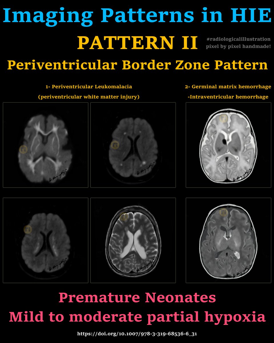
Imaging Patterns in HIE ♦️ PATTERN II Periventricular Border Zone Pattern 1- Periventricular Leukomalacia 2- Germinal matrix hemorrhage/Intraventricular hemorrhage ✔️ Premature Neonates ✔️ Mild to moderate partial hypoxia doi.org/10.1007/978-3-…


Teaching point (case summary): In cases showing mucus plugging, bronchiectasis (or peribronchial thickening), and tree-in-bud opacities, particularly with a history of recurrent chest infections, the radiologic differential can be approached based on lobar predominance. •
26-year-old male with recurrent chest infections. HRCT video shared. 🔍 Question for radiologists: • What are the key HRCT findings? • Based on imaging, what is your differential diagnosis for the underlying cause? Think beyond infection alone — consider structural and

May I introduce you to the hippocampus? Bilateral stimulation of the temporal lobes within which it is located generates the "God Experience". journals.sagepub.com/doi/abs/10.246…


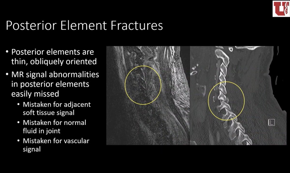
Some significant C-spine injuries are occult on MRI. Key example is fracture of the posterior elements, which have little marrow with which to show edema signal. @lubdha_shah on cervical spine misses at #RSNA2025
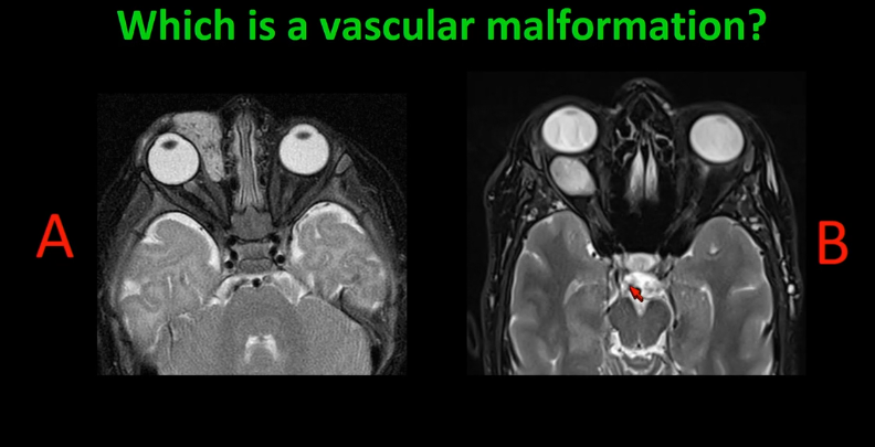
Hemangiomas are moderately T2 bright and may have flow voids. Venous malformations (formerly/incorrectly known as cavernous hemangiomas) are very T2 bright and don't have flow voids. @DShatzkes at #RSNA25
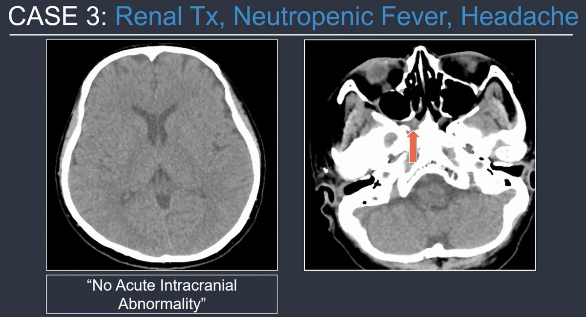
Immunocompromised patient, headache, head or face CT. You see sphenoid mucosal thickening. Scrutinize the fat spaces. 👀 This was a case of invasive fungal sinusitis. The right pterygopalatine fossa is infiltrated. 🚩 I've seen this same miss! @tabby_kennedy at #RSNA25

This guy literally breaks down how to find your life’s purpose


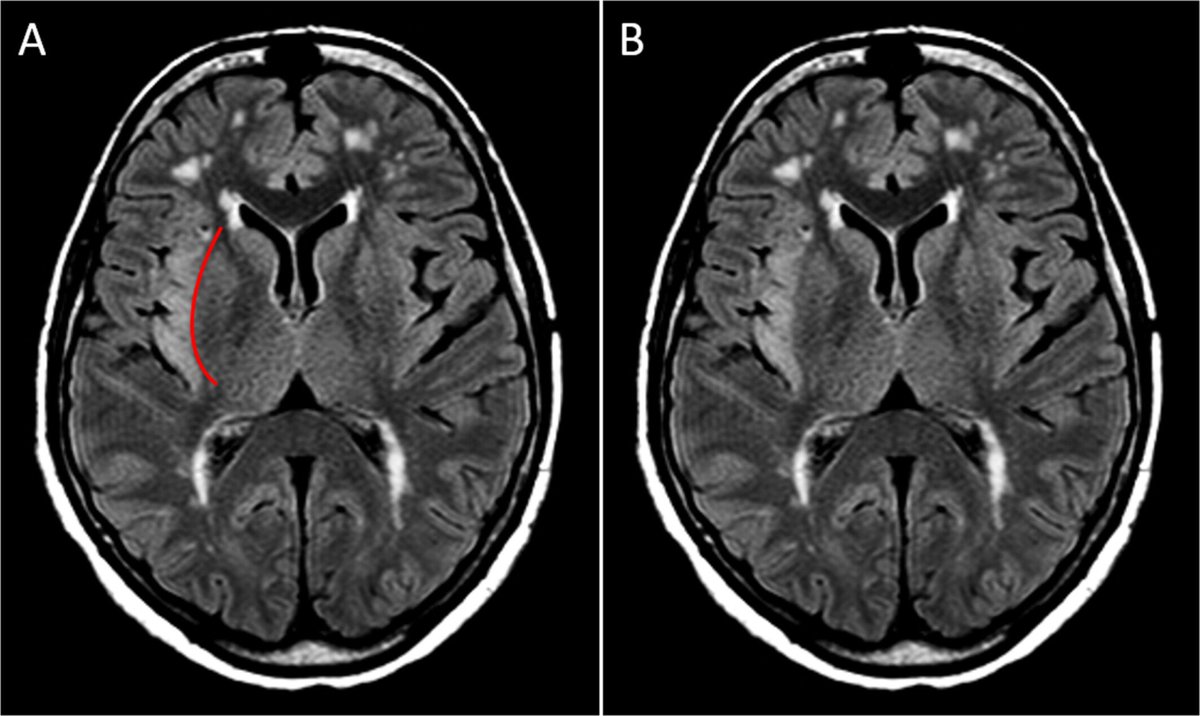
Delayed Post-Hypoxic Leukoencephalopathy (DPHL). Acute hypoxia → diffusion restriction in cortex, basal ganglia, hippocampi. First set of images (DWI and FLAIR) 2-4 weeks later → cortex normalizes, but white matter turns bright on FLAIR = DPHL (the delayed sequel of


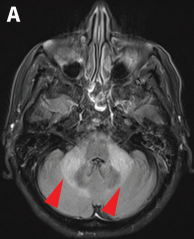
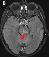
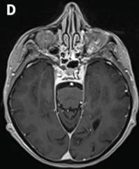
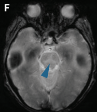
✅Rhombencephalitis due to JC virus, a form of progressive multifocal leukoencephalopathy. 💡In immunocompromised patients, especially with CLL, JC virus may involve the brainstem and cerebellar peduncles. Think of PML when lesions show no enhancement. doi.org/10.1503/cmaj.2…

🧑🦳An 86-year-old woman with long-standing chronic lymphocytic leukemia developed progressive gait imbalance, dysarthria, and binocular diplopia over 4 weeks. No fever or infection signs. 🥛CSF: mild lymphocytic pleocytosis, ↑protein, normal glucose. 🧑⚕️What’s the cause?
💡In glioblastoma, restricted diffusion outside the enhancing tumor (IRD) often heralds future enhancement. 📰Prospective cohort n=52: IRD in 21%; 64% enhanced on follow-up at ~110 days; IRD linked to higher mortality risk (HR≈3.2). doi.org/10.1007/s00234…


The “insular knife-cut” sign: a sharp FLAIR demarcation between insular hyperintensity and spared basal ganglia. Found in 70% of HSV encephalitis cases and highly specific (99%). Its presence strongly predicts HSVE even when PCR is pending or negative. doi.org/10.1111/ene.70…
Hopkins criteria for response assessment in head and neck cancer: radiopaedia.org/articles/head-… pubmed.ncbi.nlm.nih.gov/24947059/

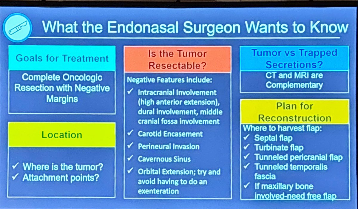
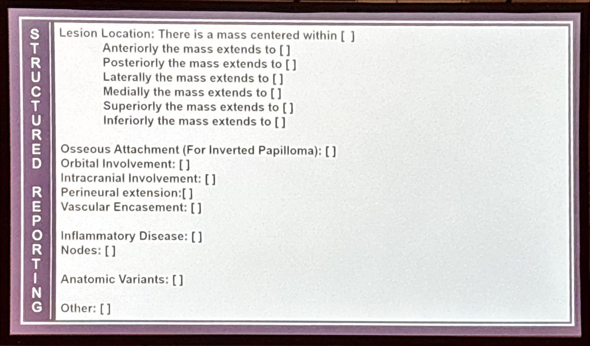
When reporting imaging of a sinonasal tumor, describing tumor extent is critical for treatment planning, less so predicting the pathology (besides concluding it is not simple inflammatory disease). @tabby_kennedy at #ASHNR25
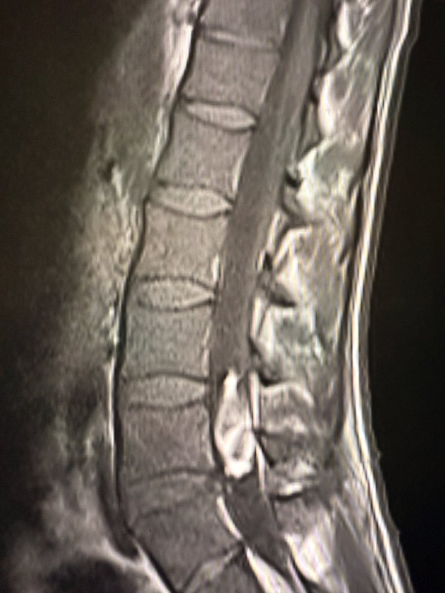
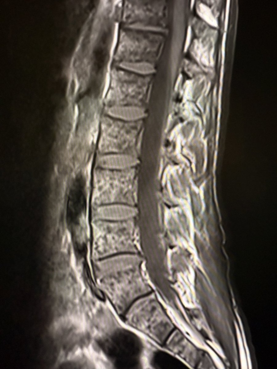
In treated leukemia, bone marrow tells its own story. Baseline (left image): There is a diffuse low signal on T1W images, and the vertebral marrow looks uniformly “dark.” The signal is lower than that of the disc. Six weeks follow-up after therapy (right image): diffuse marrow

Creating a daily routine to remain updated in your medical speciality using PubMed: PubMed is a goldmine of medical literature,updated daily By creating email alerts in PubMed for your medical specialty's journals, you get new article updates in your inbox every day A how-to🧵

Normal marrow> disc > muscle on T1W MRI. If marrow < disc: think pathology (infiltration, reconversion, anemia, storage disease). If marrow < muscle: infiltration is very likely. Differential Checklist (Diffuse Low T1 Marrow Signal): 1. Malignant infiltration (leukemia,


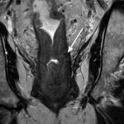
Vesicourachal diverticulum. 10yo F.

A flattened IVC defined as a transverse to antero-posterior diameter ratio of 3/1 or more seen at multiple levels has been associated with significant hypotension and / or impending shock in trauma patients. A surrounding hypodense halo as well as small caliber of the abdominal


United States Trends
- 1. Kash N/A
- 2. Tourette N/A
- 3. #AKOTSK N/A
- 4. Claressa N/A
- 5. #LCDLF6 N/A
- 6. Lakers N/A
- 7. FBI Director N/A
- 8. #RHOP N/A
- 9. #IndustryHBO N/A
- 10. #AKnightOfTheSevenKingdoms N/A
- 11. 960 SAT N/A
- 12. Pritchard N/A
- 13. Maekar N/A
- 14. El Mencho N/A
- 15. Celtics N/A
- 16. Oriana N/A
- 17. Kelly Price N/A
- 18. Canadians N/A
- 19. México N/A
- 20. Horacio N/A


Something went wrong.
Something went wrong.