Vous pourriez aimer


1st European Joint Meeting of the International Society of Dermatopathology #ISDP #dermatopathology Dr. Kempf on CD8 lymphoproliferative disorders

1st European Joint Meeting of the International Society of Dermatopathology program #ISDP #dermatopathology

1st European Joint Meeting of the International Society of Dermatopathology an European Society of Pathology starts today in Vienna. First talk is by Dr. Fraitag #ISDP #dermatopathology

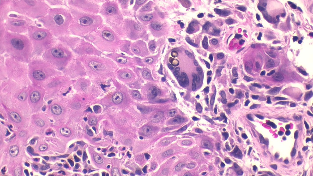
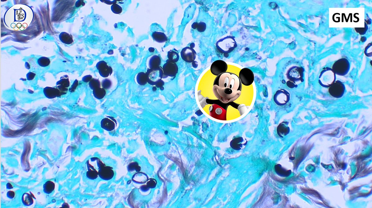
#ISDP #Olympics Spot diagnosis Case 20 - @JCandidoXavier CHROMOBLASTOMYCOSIS •Pigmented (dematiaceous) fungi •Prominent pseudoepitheliomatous hyperplasia •Granulomatous infiltrates with round brownish Medlar bodies resembling copper pennies or coffee beans #path #derm
#ISDP #Olympics Spot diagnosis Case 19 - @JoyceSSLee SEBACEOUS CARCINOMA • Periocular (75%) • Basophilic germinative sebaceous cells • Sebocytes with vacuolated cytoplasm • Nuclear pleomorphism • Increased and atypical mitoses • Necrosis #Path #derm #pathology


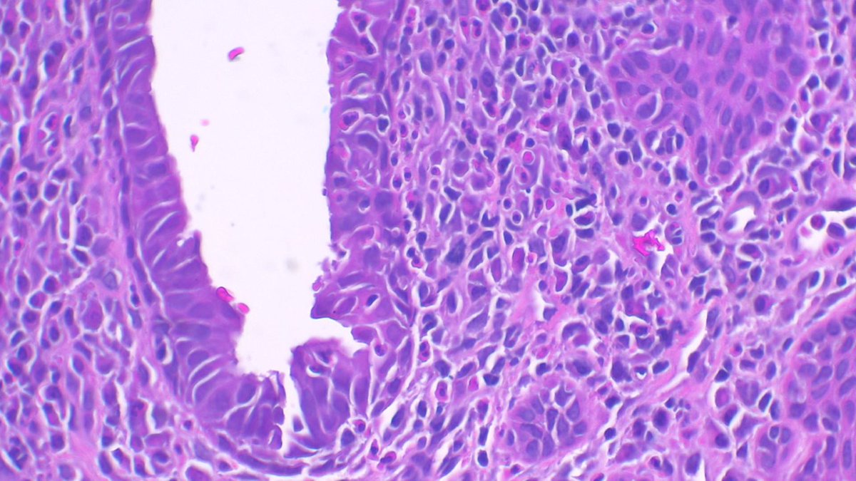
#ISDP #OlympicGames Spot diagnosis Case 18 - @JCandidoXavier SYRINGOCYSTADENOMA PAPILLIFERUM •It can occur in naevus sebaceus of Jadassohn •Endophytic crateriform lesion •Two layers of cells forming papillary and cystic structures • Numerous plasma cells #path #derm


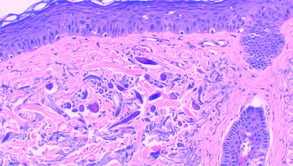
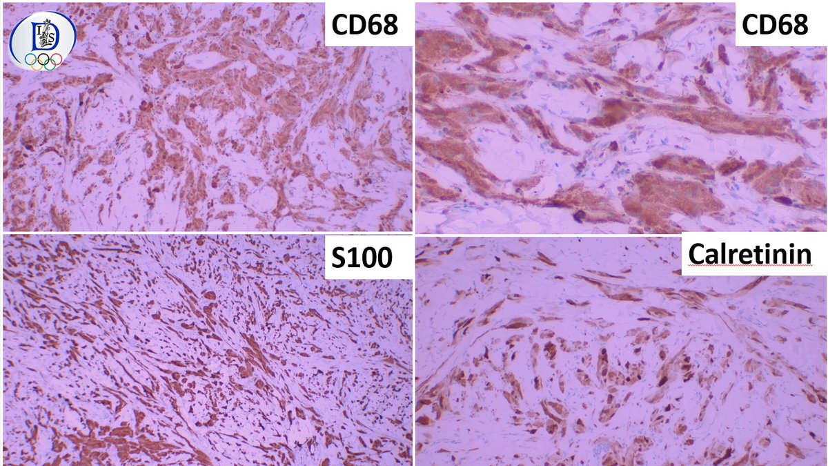
#ISDP #OlympicGames Spot diagnosis Case 17 - @JoyceSSLee CUTANEOUS ROSAI DORFMAN DISEASE •Non-Langerhans cell histiocytosis •Large histiocytes exhibiting emperipolesis (containing inflammatory cells) •CD68+, S100+, CD1a- •Lymphoplasmacytic infiltrate #Path #derm


#ISDP #Olympics Spot diagnosis Case 16 - @JCandidoXavier ELASTOFIBROMA •Benign soft tissue tumor characterized by abnormal elastic fibres and fibroblasts •Hypocellular tumor with fragmented and thickened to globular elastic fibres #Pathology #path #dermatology #derm

#ISDP #OlympicGames Spot diagnosis Case 15 - @JoyceSSLee PLEVA: •Parakeratosis •Apoptotic keratinocytes •Basal vacuolar alteration •Dermal and intraepidermal hemorrhage •Wedge-shaped lymphohistiocytic infiltrate •+/- Lymphocytic vasculitis #derm #path #pathology


#ISDP #OlympicGames Spot diagnosis Case 14 - @JCandidoXavier PARACOCCIDIOIDOMYCOSIS •Dimorphic fungi (mycelial in the environment and yeast in the host) •Granulomatous reaction pattern •Round yeast with single or multiple buds with narrow necks. #derm #path #pathology
#ISDP #OlympicGames Spot diagnosis Case 13 - @JoyceSSLee ONYCHOMATRICOMA •Thickened nail plate •Wormwood-like or honeycomb-like cavities •Papillary digitate projections covered by matrix epithelium •V-shaped keratogenouszones •Fibrocellular stroma #path #dermatology


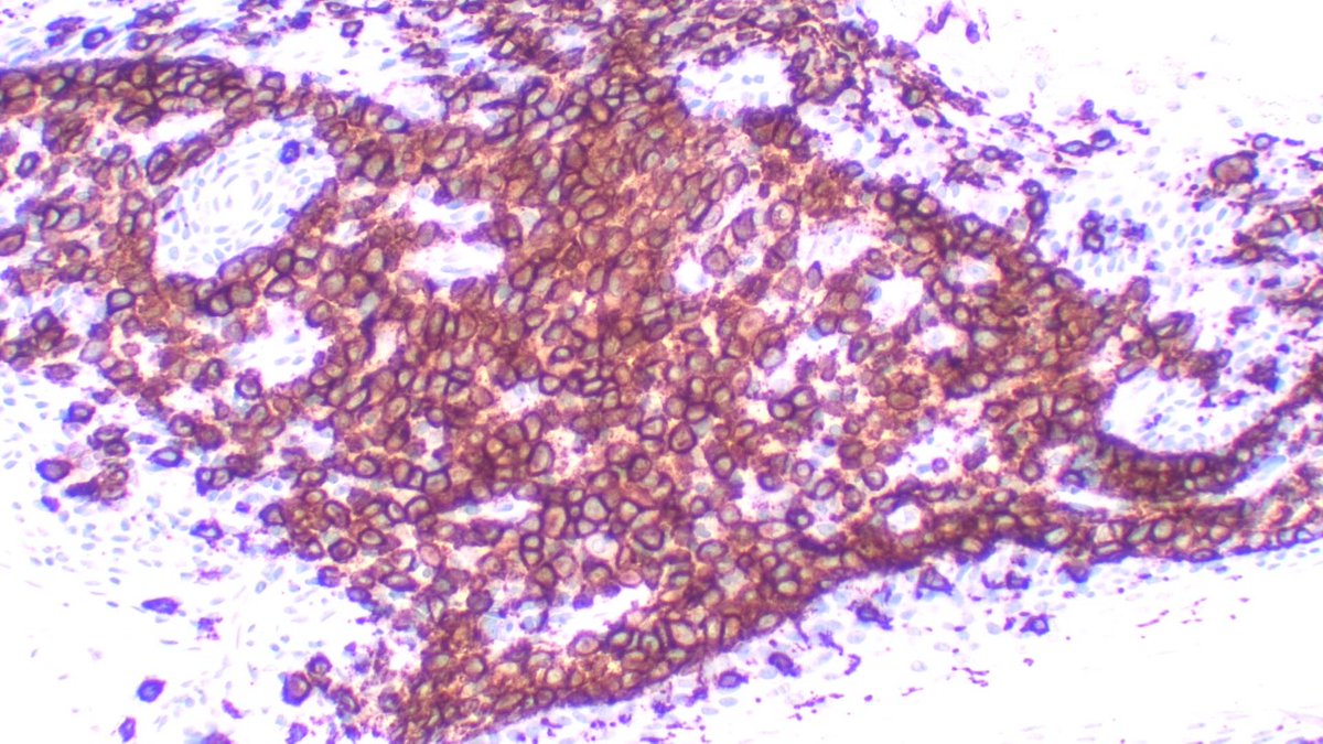
#ISDP #OlympicGames Spot diagnosis Case 12 - @JCandidoXavier LANGERHANS CELL HISTIOCYTOSIS • Round/ovoid medium-sized cells with indented, lobulated or coffee-bean nuclei. • Abundant and eosinophilic cytoplasm •IHQ markers: CD1a, Langerin and S100 protein #derm #path


Join us for three days in Queenstown, New Zealand for insightful discussions, engaging presentations, and networking opportunities with leading experts and professionals in the field of Dermatopathology. #ISDP #Dermpath #path #pathology @JMGardnerMD @GeronimoJrLapac
#ISDP #Olympics Spot diagnosis Case 11 - @JoyceSSLee NECROBIOTIC XANTHOGRANULOMA •Areas of necrobiosis •Xanthogranulomatousinflammation (foamy histiocytes, Touton giant cells) •Cholesterol clefts •Large, bizarre angulated giant cells with many nuclei #PathTwitter #derm


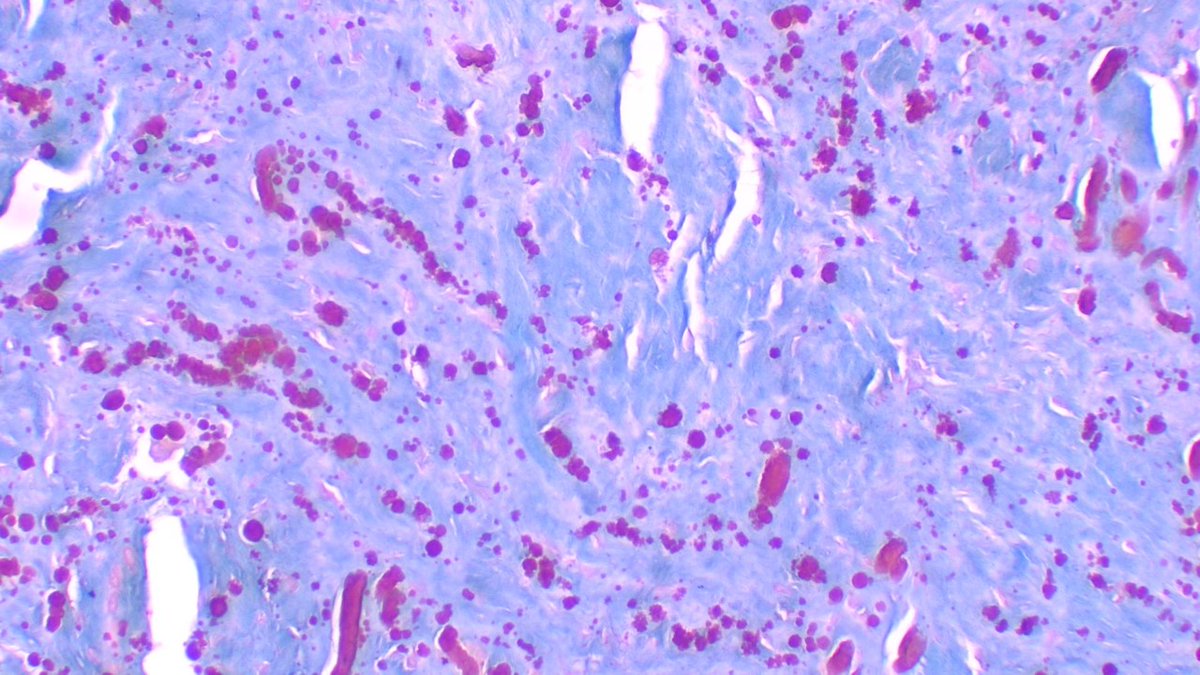
#ISDP #Olympics Spot diagnosis Case 10 - @JCandidoXavier PSEUDOXANTHOMA ELASTICUM • degenerative disease of elastic tissue • autosomal dominant • Elastic fibers are basophilic and irregular, appearing as widely dispersed granular material. #Path #dermpath #derm


#ISDP #Olympics2024 Spot diagnosis Case 9 - @JoyceSSLee NECROTIZING INFUNDIBULAR CRYSTALLINE FOLLICULITIS •Folliculocentric crater •Eosinophilic filamentous material •Material likely derived from Malassezia yeasts, bacteria, and/or sebaceous lipids #path #dermatology


#ISDP #Olympics Spot diagnosis Case 8 - @JCandidoXavier OCHRONOSIS •Clinical presentation: blue–black cutaneous pigmentation •Typical swollen, irregular, golden brown banana 🍌shaped, sickled or round ochronotic bodies #dermpath #dermatology #pathology #path

#ISDP #Olympics Spot diagnosis Case 7 - @JoyceSSLee HAND, FOOT AND MOUTH DISEASE •Caused by coxsackievirus A16, enterovirus 71 •Intraepidermal blister •Ballooning and reticular degeneration •Epidermal necrosis •Neutrophils #Dermatology #derm #dempath #pathology


#ISDP #olympics Spot diagnosis Case 6 - @JCandidoXavier GRANULAR CELL TUMOR •Benign neuroectodermal tumor •Large cells with abundant and granular cytoplasm •Synonym: Abrikossoff tumor •Pseudoepitheliomatous hyperplasia can be prominent #path #pathology #dermatology

#ISDP #Olympics Spot diagnosis Case 5 - @JoyceSSLee SCLEROMYXEDEMA •Haphazard proliferation of fibroblasts within the reticular dermis •Fibrosis and skin thickening •Increased dermal mucin #dermatology #dermatopatology #dermapath #path


United States Tendances
- 1. Blue Origin 7,124 posts
- 2. Megyn Kelly 29.3K posts
- 3. New Glenn 8,053 posts
- 4. Vine 33K posts
- 5. Senator Fetterman 17.8K posts
- 6. CarPlay 4,332 posts
- 7. #NXXT_JPMorgan N/A
- 8. World Cup 100K posts
- 9. Brainiac 4,441 posts
- 10. Portugal 61.6K posts
- 11. Matt Gaetz 13.2K posts
- 12. Padres 29.6K posts
- 13. GeForce Season 1,017 posts
- 14. Black Mirror 5,192 posts
- 15. Eric Swalwell 25.2K posts
- 16. Katie Couric 9,472 posts
- 17. Osimhen 99.3K posts
- 18. Cynthia 112K posts
- 19. Man of Tomorrow 5,290 posts
- 20. #WorldKindnessDay 16.8K posts
Vous pourriez aimer
-
 American Society of Dermatopathology
@ASDPTweets -
 Dermpedia Courses
@Dermpedia -
 Artur Zembowicz M.D.
@DrZembowicz -
 Silvija Gottesman MD
@SGottesmanMD -
 Raj Singh MD
@mydermpath -
 Jonhan Ho
@forthejon -
 Steven
@STEVENKOLKERMD -
 Dermpath-L
@Dermpathl -
 Jisun Cha MD FAAD
@sunpungi -
 Rosalynn Nazarian MD
@RosNazarianMD -
 Daniel Skipper
@DCSkipperDO -
 Gonzalo De Toro
@gonzadetoro -
 Joseph Susa, DO
@CutisViaLux
Something went wrong.
Something went wrong.