#em_monday search results


📢 @brkornum @CarmenFalcone6 @Mishra_CBF_Lab @Neurotweeps @PBNeuroVascLab @BoasDavid @agjedde @cathnaledi @CFIN_AU @hmru_cph @VibekeHjortlund and of course @HaniehFalahati and #EM_Monday.

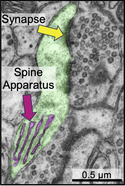
Our paper is out in time for #EM_Monday! It is about a specialized neuron-specific ER, the spine apparatus, that is close to synapses and has a peculiar shape. If you think neurons are special, the spine apparatus is their🦄horn! But how does it form?!(1/9)doi.org/10.1016/j.cub.…
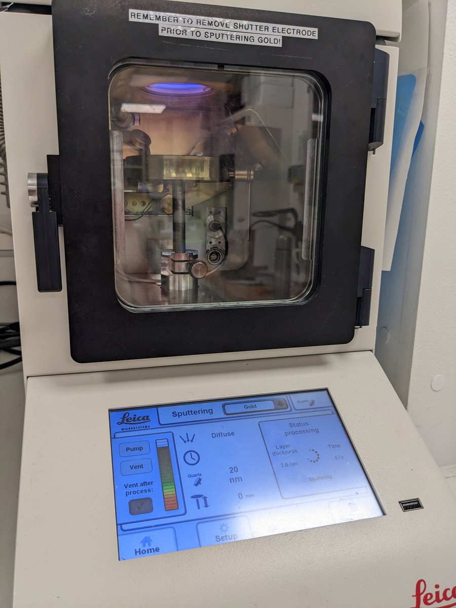
Gold plating in process! 😁 Stay tuned for more EM data. #ElectronMicroscopy #neuroscience #EM_monday #CFIM
#EM_Monday again! Ever wondered about myelination? Here is an axon in magenta as it enters the myelin sheath. As you can see, soon after it enters the sheath, myelin becomes dense being mostly membrane, and you can barely see any cytoplasm, except for some dark islands (1/n).
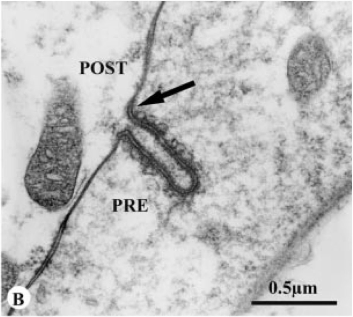
A Nowruz special of #EM_Monday!I will share what I have found about the evolution of dendritic spine. The earliest thing I found distantly resembling a spine is in the retina of box jellyfish, which shows an invagination of postsynapse into presynapse (doi.org/10.1086/bblv21…)1/n
For a belated #EM_Monday, I will share my favorite thing in neurons, the spine apparatus of a pyramidal neuron! The bulk of my postdoctoral work has been on this peculiar organelle. youtu.be/LleCdCtUnLU
Can anyone guess what this cell is that is SO densely paced with mitochondria?! #EM_Monday.Image from openorganelle.janelia.org @HHMIJanelia I was definitely not expecting this! You can see the answer and more images below.

This #EM_Monday I want to tweet about organelles that are very mysterious things to me, but have something in common: layers upon layers of membranes. I will share some and would love to see other examples that you might know of!The first example is lamellar bodies in lungs (1/n)

I think this bacteriophage negative stain is a bit scary! 🎃💀🖤🦠 #HappyHalloween #EM_monday #CFIM #EMcourse


😍 " moment of the entry of Lipofectamines into the cell via endosome-rupture" So many wonderful cell biology ideas behind how John Heuser captured these rare, transient, asynchronous moments. Read them here: early #EM_Monday @HaniehFalahati biorxiv.org/content/10.110…


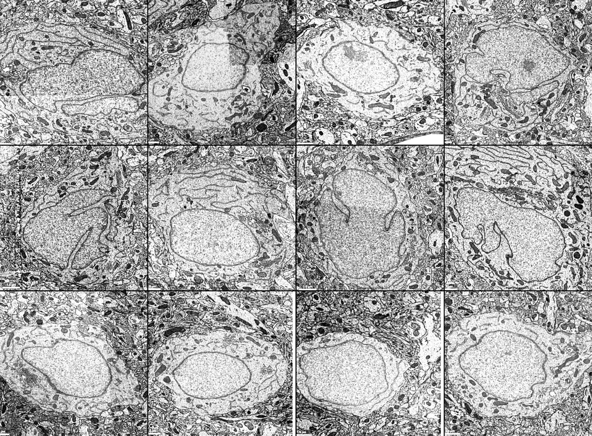
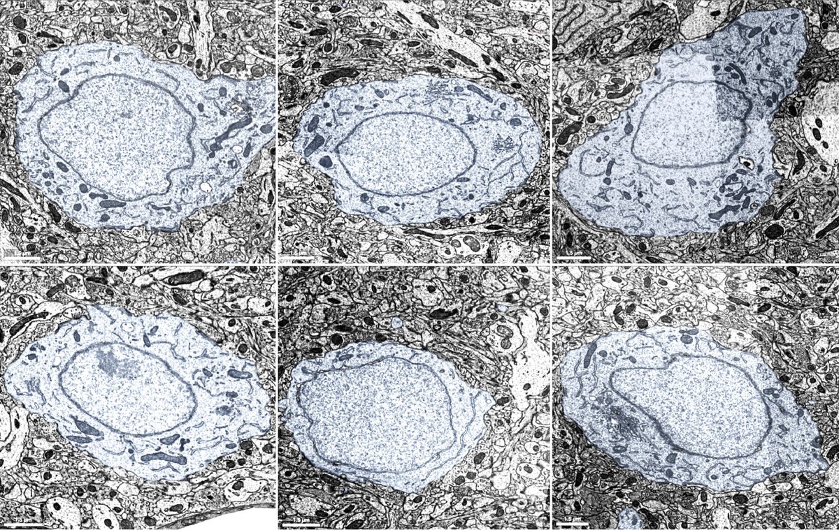
#EM_Monday again! These are randomly selected cell bodies, close to the nucleus periphery, of 6 inhibitory and 6 excitatory neurons from layer 2/3 of microns-explorer.com. Can you tell which are which?
Here comes the answers to the #MICrONS EM challenge... dum dum DUUUM! #EM_monday
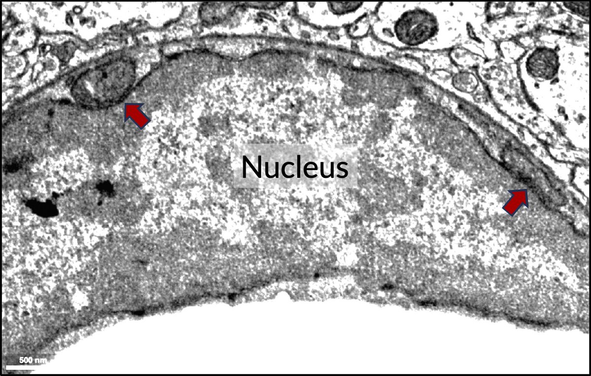
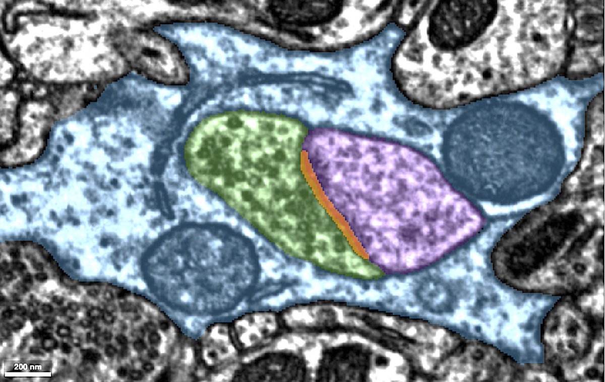
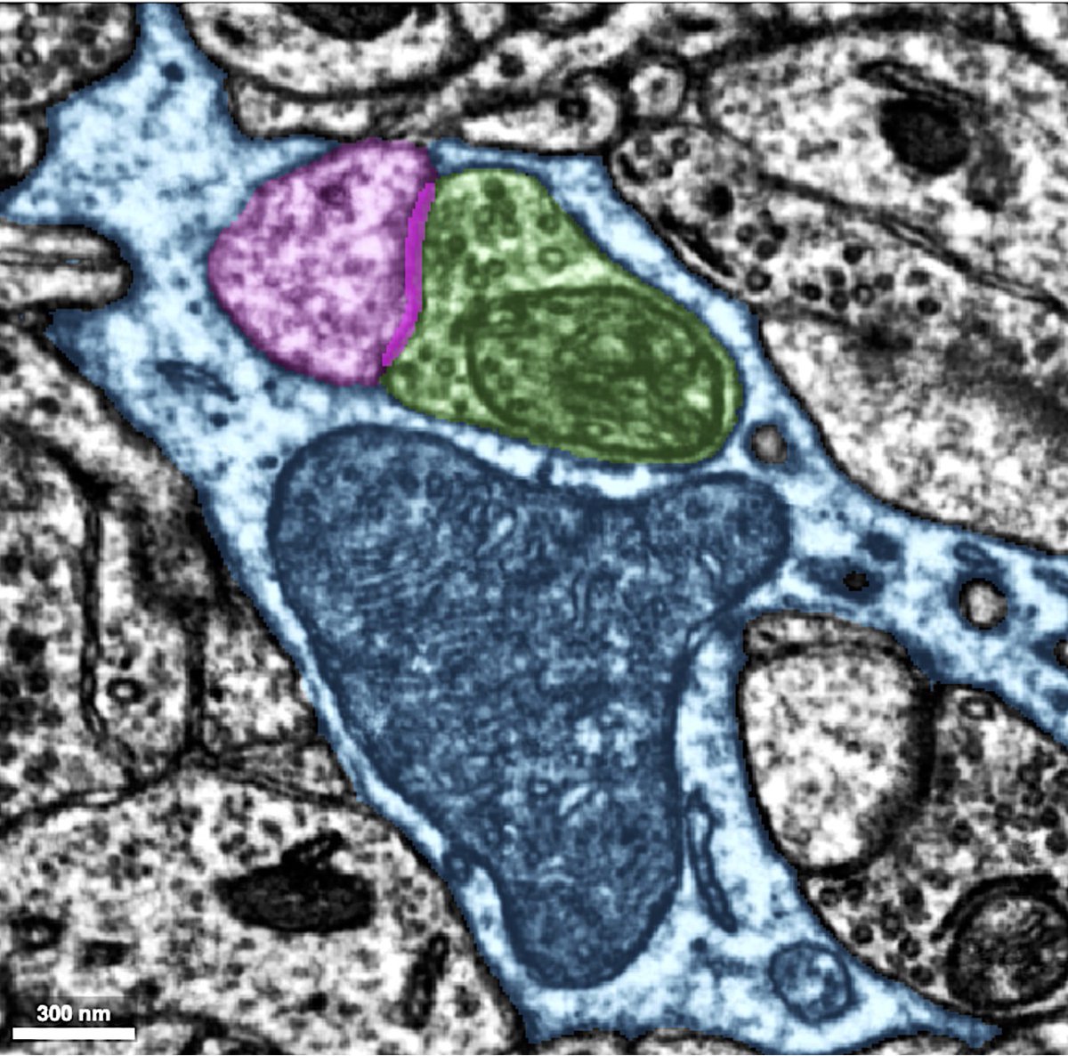
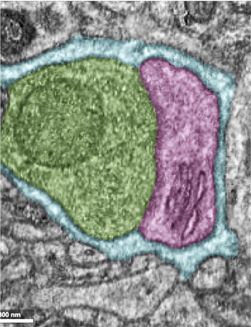
This #EM_Monday is about mitochondria-nuclear envelope contacts in different brain cells. There are many reasons for mito and NE to want to have direct lines of communications: from coordinating response to cellular stress to transport of lipids and even proteins. However, (1/n)
This #EM_Monday I share a🧵on our new paper @PDCLab. Whether you are into organelles, cytoskeleton, neurons, or just enjoy a fun story, this is for you! Organelles like ER in specialized cells can take weird shapes, w/ the spine apparatus(SA) of dendrites as a prime example (1/n)
My new paper is now on BioRxiv 🎉🎉🎉🎉 twittorial to come 😊 Ectopic Reconstitution of a Spine-Apparatus Like Structure Provides Insight into Mechanisms Underlying Its Formation biorxiv.org/content/10.110…
Look at these beautiful OPCs in action 😍 #EM_Monday

Did you know that OPCs are the #1 dividing cell in the brain? Here OPC 1 has half of the division while OPC 2 is migrating away from the pyramidal cell that host OPC1. You’re on your own buddy!

How many cell bodies can a single soma hug at the same time?! Well, for this fly neuron it seems like 9 is the magic number! #EM_Monday (1/n)

This weeks #EM_Monday is for people who like microglia, nucleus/nucleophagy(?), and mitochodria! A 🧵 In this image you are seeing a microglia (pink) near the soma of a neuron (blue). Even from this image you can see how flat the soma and nucleus of the microglia are. (1/n)

Our paper is out in time for #EM_Monday! It is about a specialized neuron-specific ER, the spine apparatus, that is close to synapses and has a peculiar shape. If you think neurons are special, the spine apparatus is their🦄horn! But how does it form?!(1/9)doi.org/10.1016/j.cub.…
This #EM_Monday I share a🧵on our new paper @PDCLab. Whether you are into organelles, cytoskeleton, neurons, or just enjoy a fun story, this is for you! Organelles like ER in specialized cells can take weird shapes, w/ the spine apparatus(SA) of dendrites as a prime example (1/n)
My new paper is now on BioRxiv 🎉🎉🎉🎉 twittorial to come 😊 Ectopic Reconstitution of a Spine-Apparatus Like Structure Provides Insight into Mechanisms Underlying Its Formation biorxiv.org/content/10.110…
📢 @brkornum @CarmenFalcone6 @Mishra_CBF_Lab @Neurotweeps @PBNeuroVascLab @BoasDavid @agjedde @cathnaledi @CFIN_AU @hmru_cph @VibekeHjortlund and of course @HaniehFalahati and #EM_Monday.
😍 " moment of the entry of Lipofectamines into the cell via endosome-rupture" So many wonderful cell biology ideas behind how John Heuser captured these rare, transient, asynchronous moments. Read them here: early #EM_Monday @HaniehFalahati biorxiv.org/content/10.110…
This weeks #EM_Monday is for people who like microglia, nucleus/nucleophagy(?), and mitochodria! A 🧵 In this image you are seeing a microglia (pink) near the soma of a neuron (blue). Even from this image you can see how flat the soma and nucleus of the microglia are. (1/n)
#EM_Monday again! Ever wondered about myelination? Here is an axon in magenta as it enters the myelin sheath. As you can see, soon after it enters the sheath, myelin becomes dense being mostly membrane, and you can barely see any cytoplasm, except for some dark islands (1/n).
After a long break, should I bring #EM_Monday back?!
40 vote · Final results
Gold plating in process! 😁 Stay tuned for more EM data. #ElectronMicroscopy #neuroscience #EM_monday #CFIM
I think this bacteriophage negative stain is a bit scary! 🎃💀🖤🦠 #HappyHalloween #EM_monday #CFIM #EMcourse
This #EM_Monday I want to tweet about organelles that are very mysterious things to me, but have something in common: layers upon layers of membranes. I will share some and would love to see other examples that you might know of!The first example is lamellar bodies in lungs (1/n)
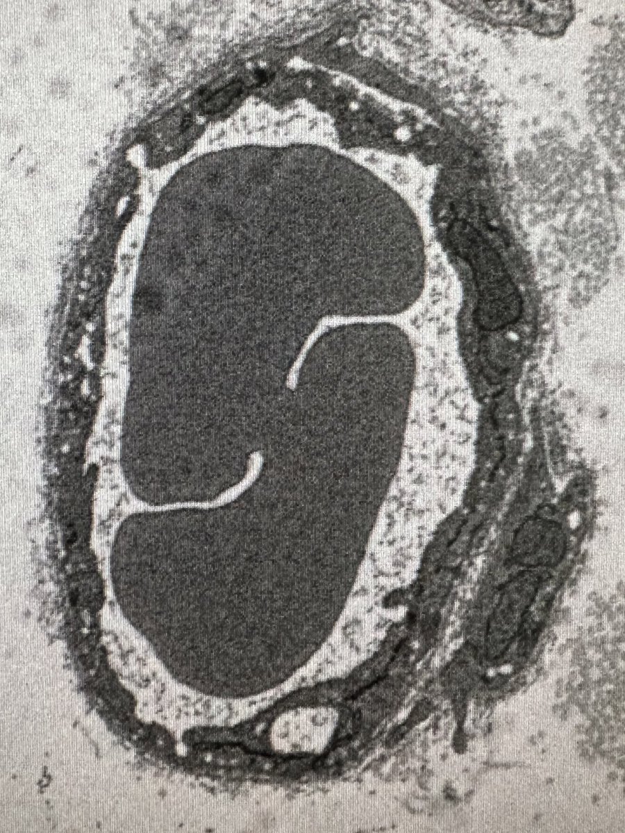
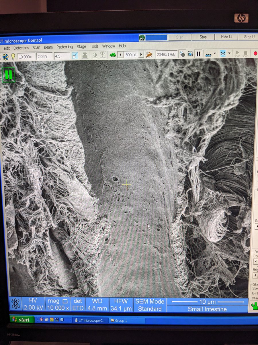
🎉 Just finished a fantastic course in electron microscopy at @UCPH_health's CFIM! 🎓 Highly recommend! Here's a thread 🧵 of some cool images we took. First up, an SEM image of a blood vessel in a small intestine. 🤩 #EM_Monday #ElectronMicroscopy
This #EM_Monday I want to share with you an image of a lipid droplet (LD) that is seemingly making contact with a mitochondria. But something is not quite right! The blue cell is a neuron, and neurons are not supposed to have LDs in normal conditions. So what is going on?! (1/n)

This #EM_Monday I want to put the word out that I will be on job market this year! As you might have noticed, I am fascinated by the shapes of various organelles that become drastically different in specialized cells. And I want to know how and why?! The most notable…(1/5)
How many cell bodies can a single soma hug at the same time?! Well, for this fly neuron it seems like 9 is the magic number! #EM_Monday (1/n)
This #EM_Monday is about mitochondria-nuclear envelope contacts in different brain cells. There are many reasons for mito and NE to want to have direct lines of communications: from coordinating response to cellular stress to transport of lipids and even proteins. However, (1/n)
Here comes the answers to the #MICrONS EM challenge... dum dum DUUUM! #EM_monday
Look at these tough swollen mitochondria of fly glial cells (red arrows), pushing their way though neuronal cell bodies, even deforming a nucleus of a neuron. And look how pathetic the neuronal mitochondria (purple arrows) look! #EM_Monday @YaleCellBio Image from @FlyWireNews

For a belated #EM_Monday, I will share my favorite thing in neurons, the spine apparatus of a pyramidal neuron! The bulk of my postdoctoral work has been on this peculiar organelle. youtu.be/LleCdCtUnLU

#NeuroArtFriday [or #EM_Monday? 😁]
Who wants to see a microglia eating a synapse for this #EM_Monday?! This is an image of a typical microglia that you find in the brain, but if you look carefully, you can see this one is hiding something in one of its hands (yellow arrow).


Can anyone guess what this cell is that is SO densely paced with mitochondria?! #EM_Monday.Image from openorganelle.janelia.org @HHMIJanelia I was definitely not expecting this! You can see the answer and more images below.
How many cell bodies can a single soma hug at the same time?! Well, for this fly neuron it seems like 9 is the magic number! #EM_Monday (1/n)
This #EM_Monday is about mitochondria-nuclear envelope contacts in different brain cells. There are many reasons for mito and NE to want to have direct lines of communications: from coordinating response to cellular stress to transport of lipids and even proteins. However, (1/n)
#EM_Monday again! These are randomly selected cell bodies, close to the nucleus periphery, of 6 inhibitory (red) and 6 excitatory (blue) neurons from layer 2/3 of microns-explorer.com. Do you notice any differences? @YaleCellBio

This #EM_Monday I will share some astrocyte hug. In these images, astrocytes (blue) surround the presynaptic boutons (green) and the dendritic spines( pink) that form a synapse.
A Nowruz special of #EM_Monday!I will share what I have found about the evolution of dendritic spine. The earliest thing I found distantly resembling a spine is in the retina of box jellyfish, which shows an invagination of postsynapse into presynapse (doi.org/10.1086/bblv21…)1/n
😍 " moment of the entry of Lipofectamines into the cell via endosome-rupture" So many wonderful cell biology ideas behind how John Heuser captured these rare, transient, asynchronous moments. Read them here: early #EM_Monday @HaniehFalahati biorxiv.org/content/10.110…
Gold plating in process! 😁 Stay tuned for more EM data. #ElectronMicroscopy #neuroscience #EM_monday #CFIM
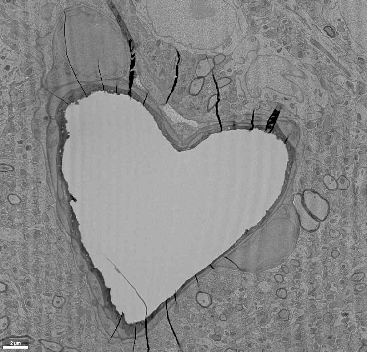
When we think about communication between neurons, we often think about synapses. But as you can see in this week's #EM_Monday images, the cell body of neurons can come into contact with each other, and this contact area is huge compared to synapses. So do the neurons use...(1/n)

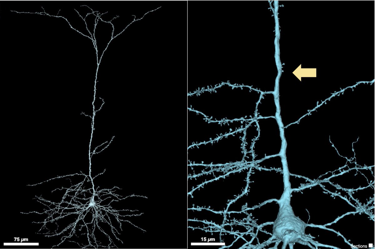
This last min #EM_Monday, I will share a beautiful pyramidal neuron I found. What was surprising to me is that for hundreds of microns, there are no dendritic spines in its apical dendrite, so for a while all synapses are onto the dendritic shaft. Arrow shows the 1st spine. (1/n)
This weeks #EM_Monday is for people who like microglia, nucleus/nucleophagy(?), and mitochodria! A 🧵 In this image you are seeing a microglia (pink) near the soma of a neuron (blue). Even from this image you can see how flat the soma and nucleus of the microglia are. (1/n)
I think this bacteriophage negative stain is a bit scary! 🎃💀🖤🦠 #HappyHalloween #EM_monday #CFIM #EMcourse
Who wants to see a microglia eating a synapse for this #EM_Monday?! This is an image of a typical microglia that you find in the brain, but if you look carefully, you can see this one is hiding something in one of its hands (yellow arrow).
Because of my PhD work, I carry this passion for membrane-less organelles. So for today's #EM_Monday I will share some of these membrane-less organelles in neurons. The first is the magnificent nucleolus, from a medium spiny neuron of songbird, showing off its subdomains.

This week's #EM_Monday is about some anomalies in neurons. The first example is at axonal terminal (green). It is often seen in Neurodegeneration, but this image but what I am showing here is from an adolescent mouse. An OPC (magenta) is touching it but there doesn't...(1/n)

Look at these tough swollen mitochondria of fly glial cells (red arrows), pushing their way though neuronal cell bodies, even deforming a nucleus of a neuron. And look how pathetic the neuronal mitochondria (purple arrows) look! #EM_Monday @YaleCellBio Image from @FlyWireNews
This #EM_Monday I share a🧵on our new paper @PDCLab. Whether you are into organelles, cytoskeleton, neurons, or just enjoy a fun story, this is for you! Organelles like ER in specialized cells can take weird shapes, w/ the spine apparatus(SA) of dendrites as a prime example (1/n)
My new paper is now on BioRxiv 🎉🎉🎉🎉 twittorial to come 😊 Ectopic Reconstitution of a Spine-Apparatus Like Structure Provides Insight into Mechanisms Underlying Its Formation biorxiv.org/content/10.110…
🎉 Just finished a fantastic course in electron microscopy at @UCPH_health's CFIM! 🎓 Highly recommend! Here's a thread 🧵 of some cool images we took. First up, an SEM image of a blood vessel in a small intestine. 🤩 #EM_Monday #ElectronMicroscopy
#EM_Monday again! These are randomly selected cell bodies, close to the nucleus periphery, of 6 inhibitory and 6 excitatory neurons from layer 2/3 of microns-explorer.com. Can you tell which are which?
Something went wrong.
Something went wrong.
United States Trends
- 1. Samara Cyn N/A
- 2. Good Friday N/A
- 3. #GuvenliLimanTurkiye N/A
- 4. Fergio House N/A
- 5. Miles Mikolas N/A
- 6. $LOL N/A
- 7. F-15 N/A
- 8. ALL RISE N/A
- 9. Tommy Lloyd N/A
- 10. Happy Easter N/A
- 11. Will Warren N/A
- 12. #FursuitFriday N/A
- 13. Eury Perez N/A
- 14. Mike Evans N/A
- 15. Grandma and Grandpa N/A
- 16. Andy Pages N/A
- 17. Fenway N/A
- 18. Xander Bogaerts N/A
- 19. CSAR N/A
- 20. #Boycott_HYBE_BELIFTLAB N/A