#electronmicroscopy search results

Adult human brains have between 86-100 billion neurons and every single one is unique - like snowflakes! ❄️ In this 3D rendering by our #ElectronMicroscopy team, we see 3 different shapes incl. (from left to right) a basket cell, tufted excitatory cell, and a Martinotti cell.
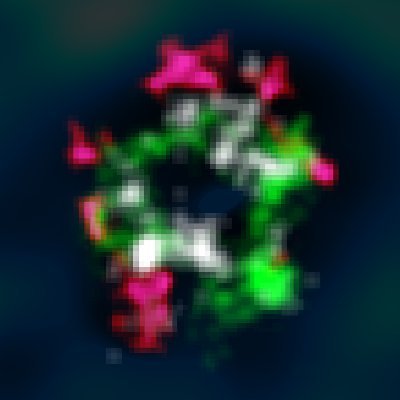
Glia derives from the greek word for glue, but they are so much than that. In this video by our #ElectronMicroscopy team, shows a pyramidal neuron (PyC) surrounded by 8 glia brain cells – 5 microglia (MG) and 3 oligodendrocyte progenitor cells (OPC). 🔗 microns-explorer.org

Happy National Cake Day ! 🎂🍰🍓 (November 26) #ElectronMicroscope #electronmicroscopy Cryo-SEM of air bubbles and fat globules in fresh cream

Just a single, happy #eosinophil patrolling the human intestine! #electronmicroscopy #MicroscopyMonday #sciart

Here's a high-resolution peek at #neutrophils isolated straight from the human bloodstream, captured in stunning detail with #electronmicroscopy and colorized! 🤩 #MicroscopyMonday


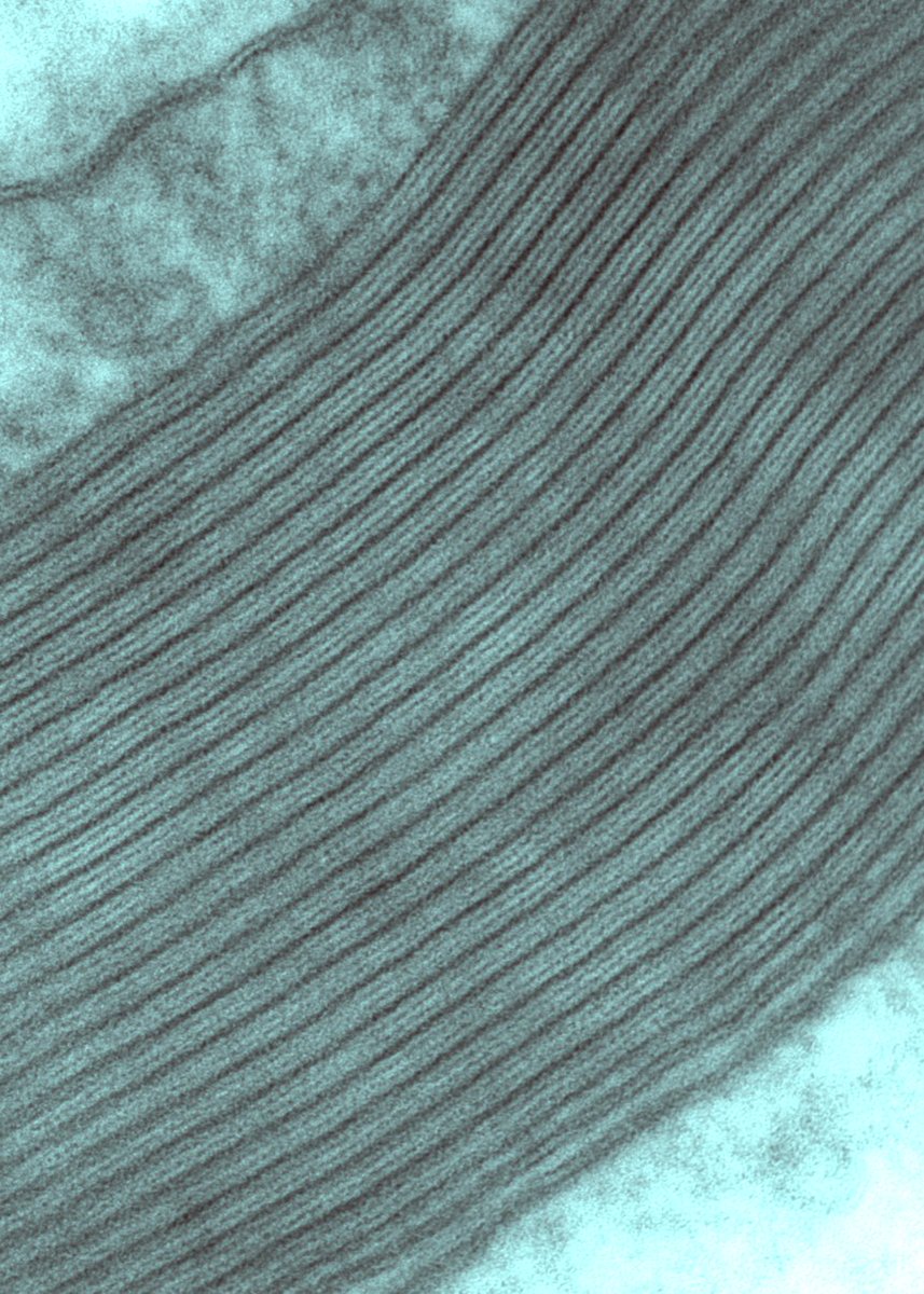
Myelin sheath is a multilamellar spiral of cell membrane around an axon, consisting of alternating major dense and two intraperiod lines. Several diseases and conditions, such as multiple sclerosis, degrade myelin compromising the optimal electrical signal. #electronmicroscopy
When the sentinel wakes! 🤩 An activated #neutrophil captured under #electronmicroscopy showing a uropod! #MicroscopyMonday #sciart

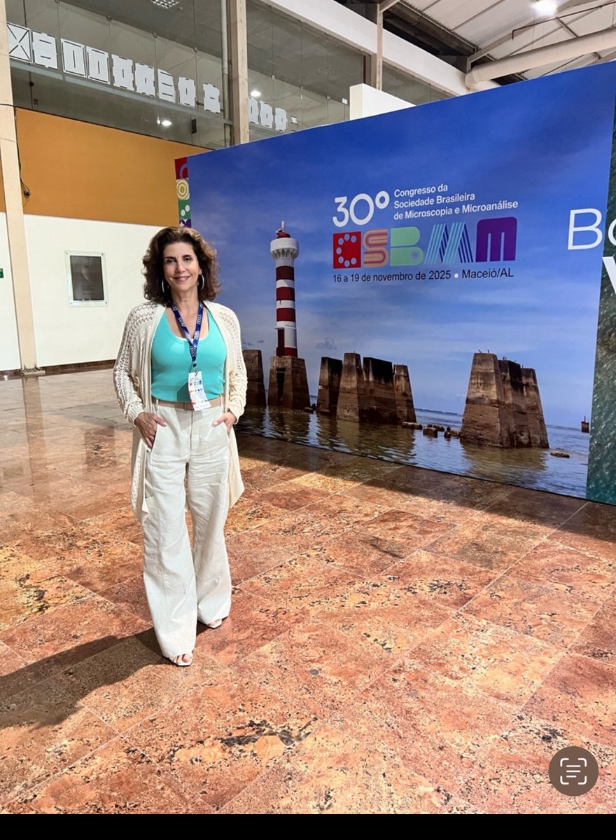
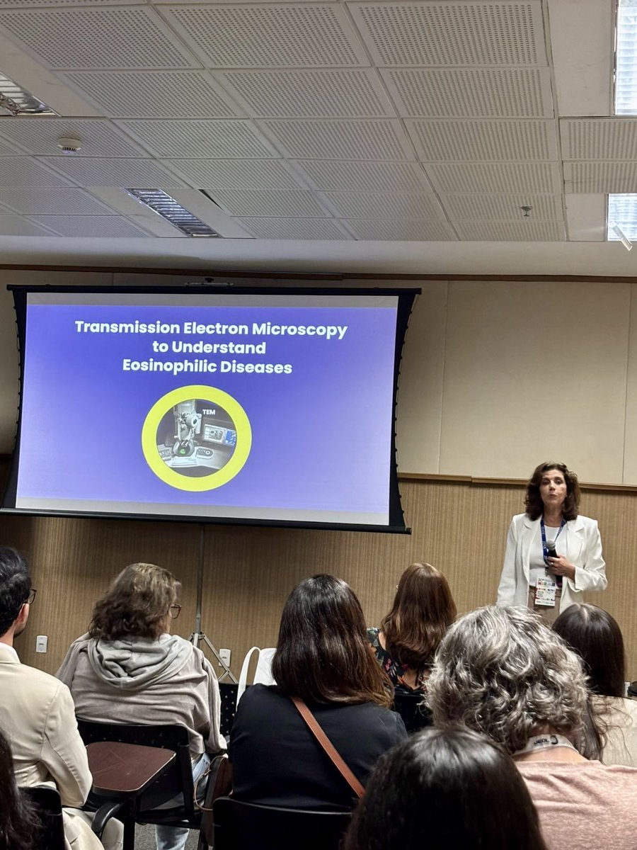
Happy and honored to give a lecture at the 30th Congress of the Brazilian Society for Microscopy and Microanalysis in beautiful Maceió! #electronmicroscopy #microscopy #WomenInScience
Neurons come in many different morphologies or shapes. Check out just some of the diversity of just inhibitory neurons in this beautiful video by our #ElectronMicroscopy team.

Starting the week with these nice membranes is one of the best things it could have happened to me. Finally happy 🥰 #ElectronMicroscopy


My in-person talk at the @GordonConf vEM meeting is right around the corner! I will present @GSegala's, @LoogerL's, and my work: new chemically resistant fluorescent proteins (& enzymes!) for #CLEM / #ElectronMicroscopy. Come say hi! #Eos5 #hfYFP (and more) at #GRC #Microscopy


Electron micrograph of axonal swellings surrounding an Aβ plaque in the cortex of a 5xFAD mouse 🧠 Note the abundance of electron-dense autophagic vesicles in these swellings. #ElectronMicroscopy


Melainia McClain, a #scientist in our #ElectronMicroscopy team, advises & assists our researchers with a wide range of 3D EM imaging techniques. #microscopy #SerialSectioning #innovation #ArrayTomography #SPOTLIGHTSTOWERS

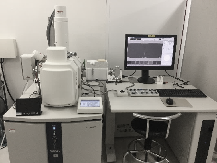
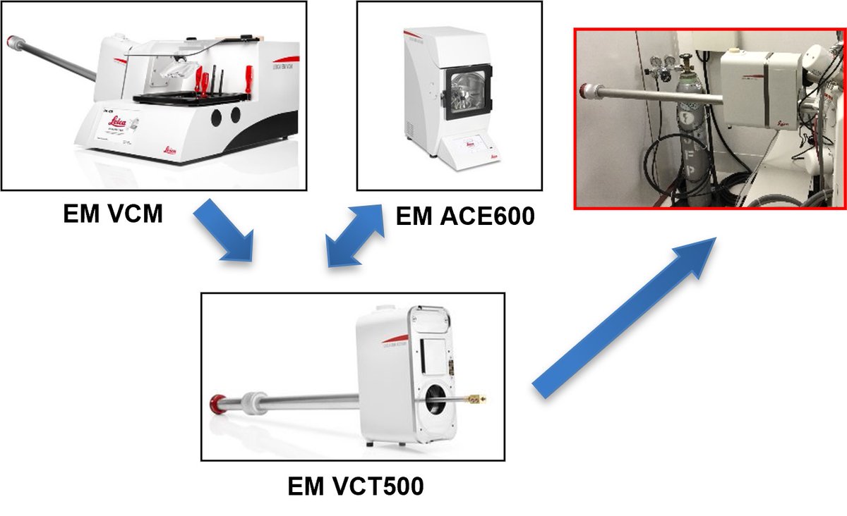
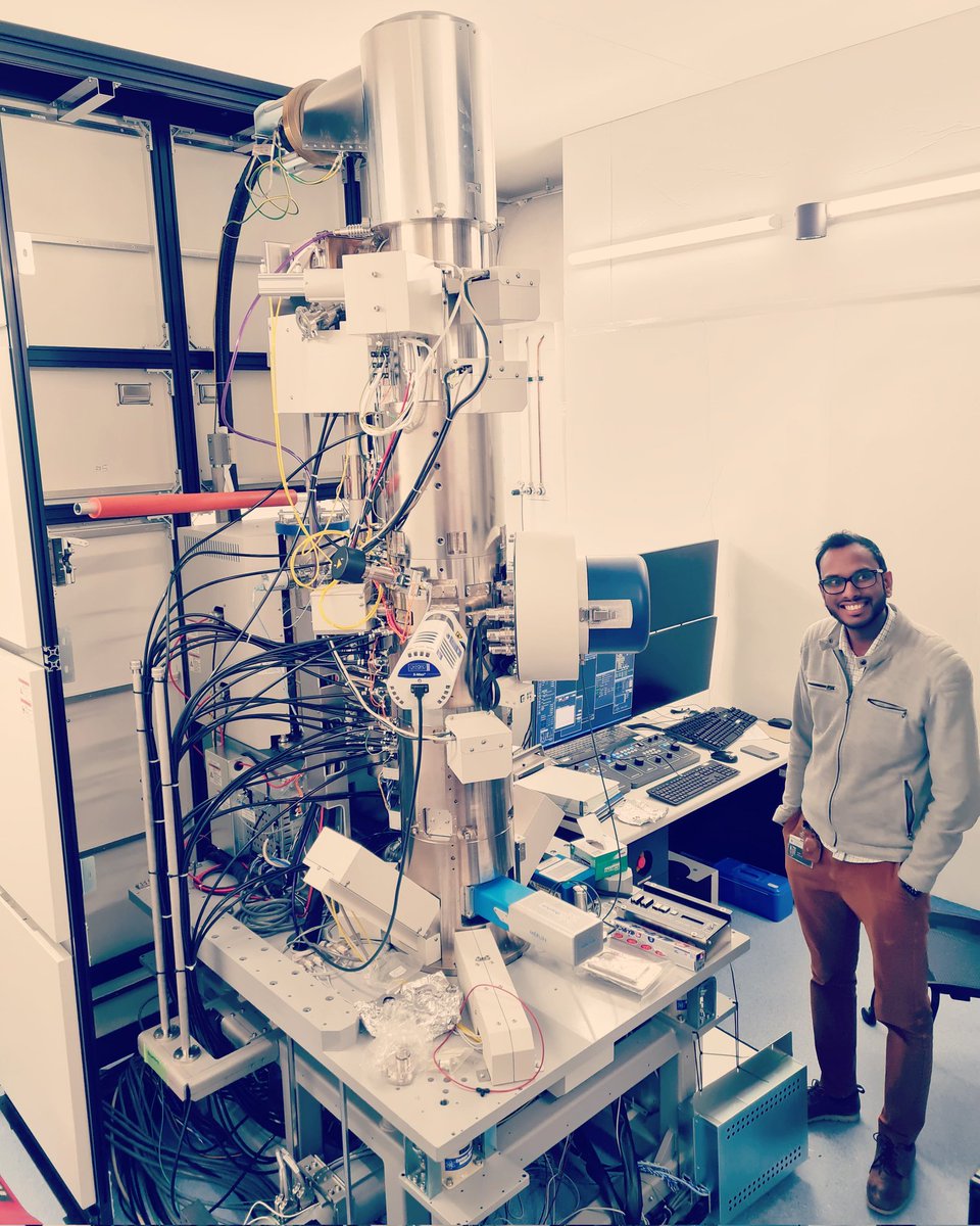
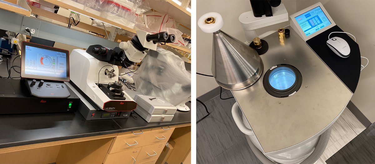
Excited to have the UC Enuity Ultramicrotome and AFS2 from @LeicaMicro! Thanks @DnbResearch and @StJudeResearch for the support! I’ve never seen the substitution chamber in a completely silver color before. #ElectronMicroscopy #NewPI

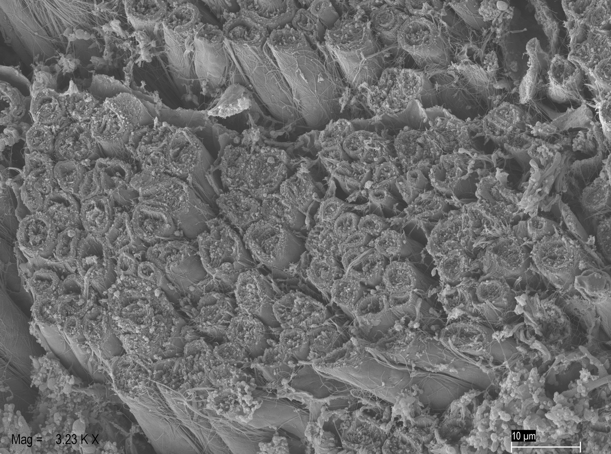
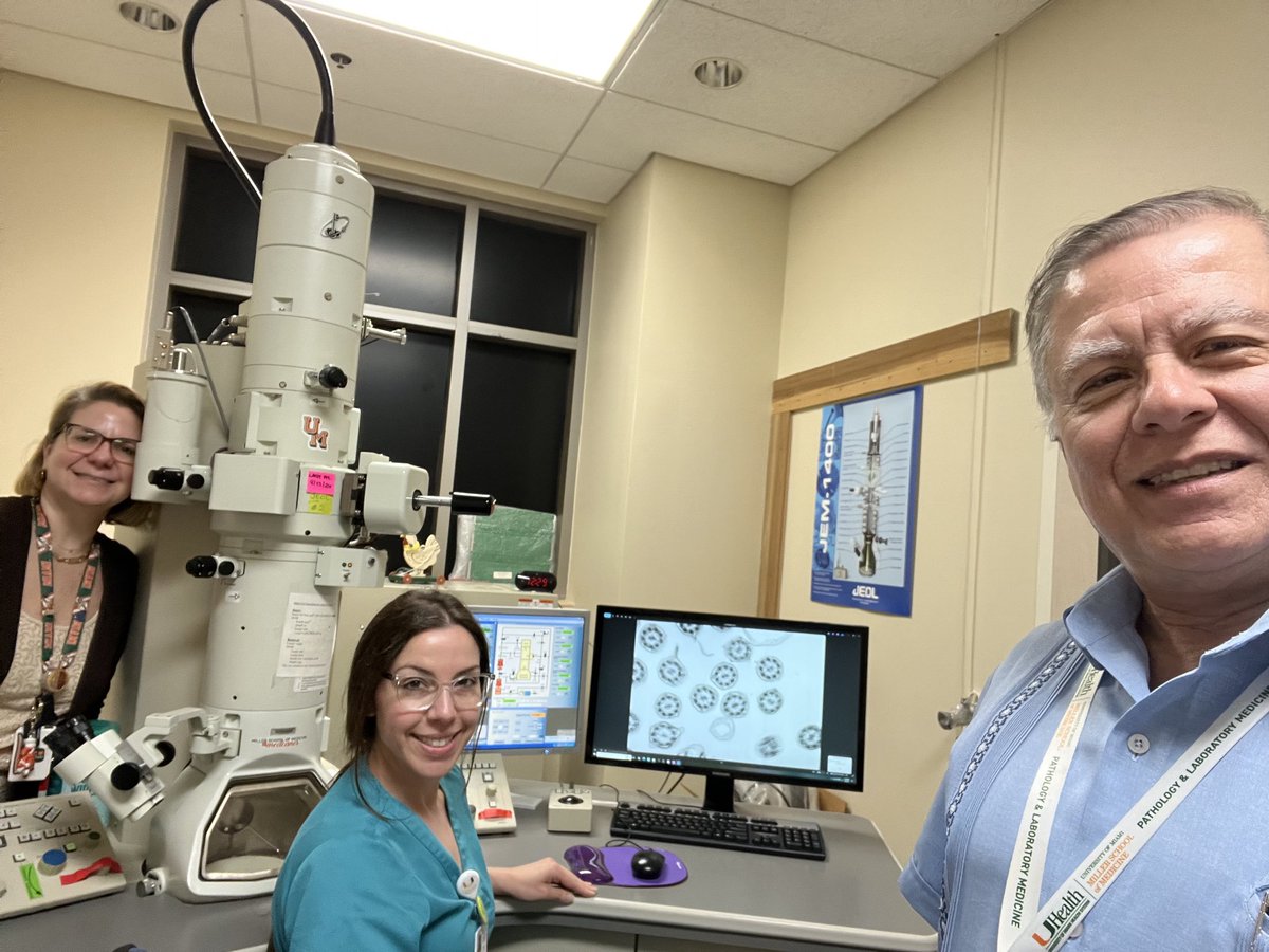
Among the many interesting reasons to do #PediPath is the “artistic science” of studying #cilia by video-microscopy and transmission #ElectronMicroscopy. Here we are with two wonderful colleagues at the EM core of the @univmiami and @UMiamiPathology

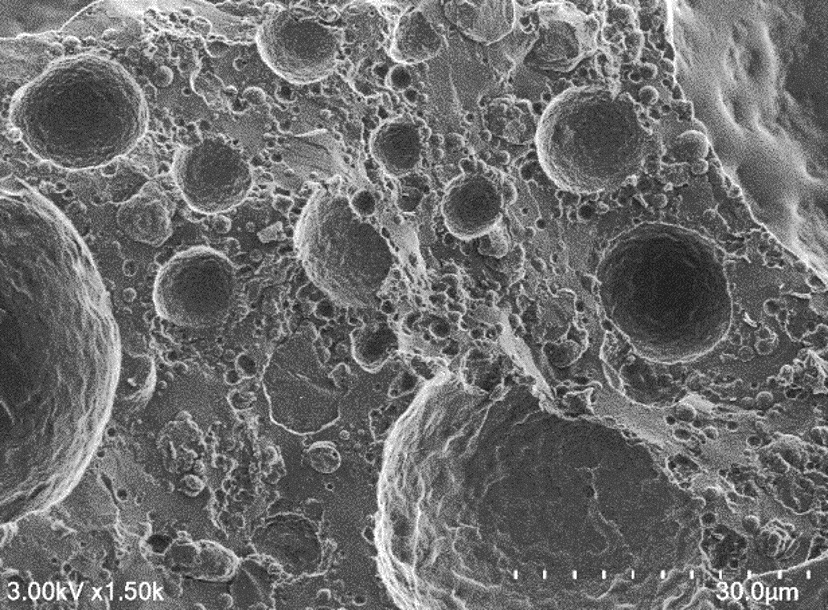
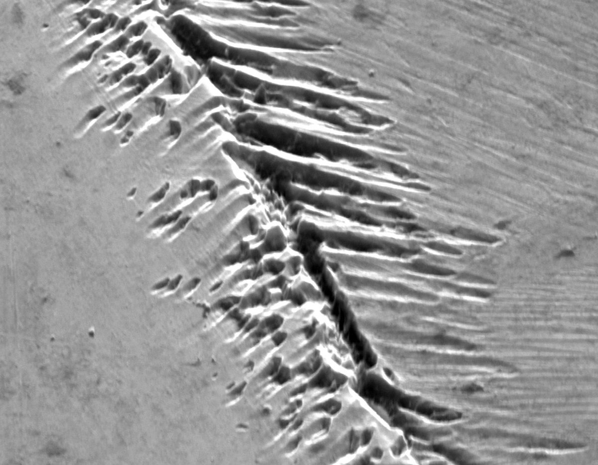
I found a mini-MOR in the SEM today. Doesn't this look like mid-ocean ridge topography? (Etched grain boundary in an alloy, field of view ca. 30 µm.) #ElectronMicroscopy
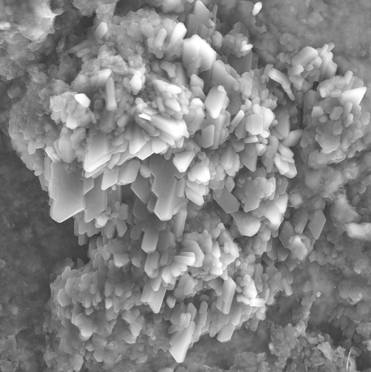
I usually prefer working with thinsections, but sometimes 3D samples are fun as well! Here some gypsum growing in black shale from Oslo. Field of view ca 10 µm. #electronmicroscopy

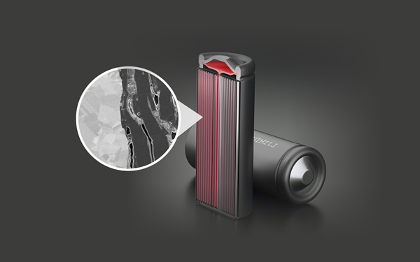
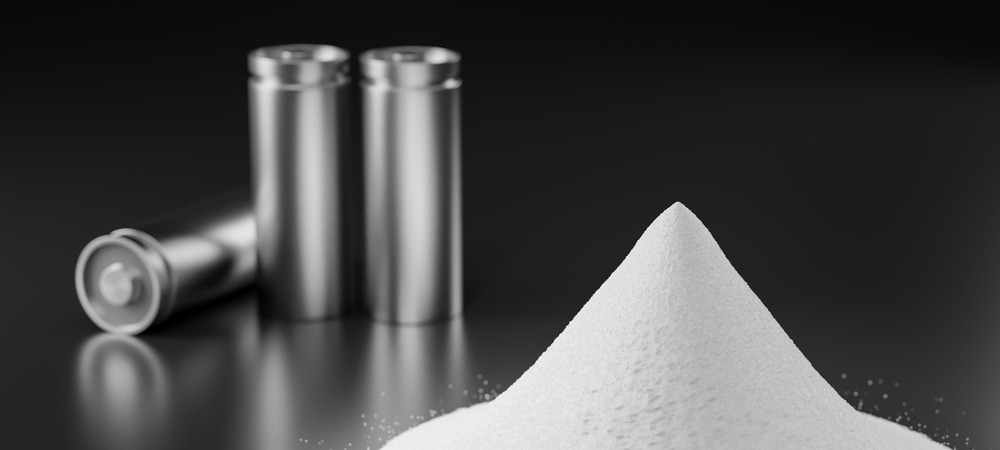
See your batteries like never before 🔬: Electron microscopy reveals secrets at the micro- and nano-scale, shaping the future of energy storage. Want to know what really drives battery performance? Dive in: azom.com/article.aspx?A… #BatteryResearch #ElectronMicroscopy ⚡

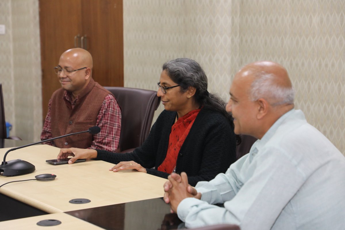
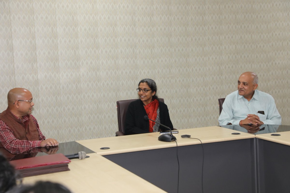
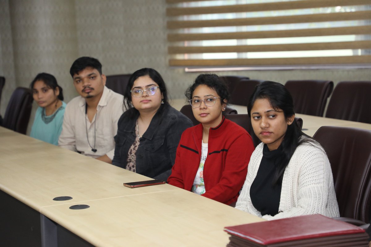
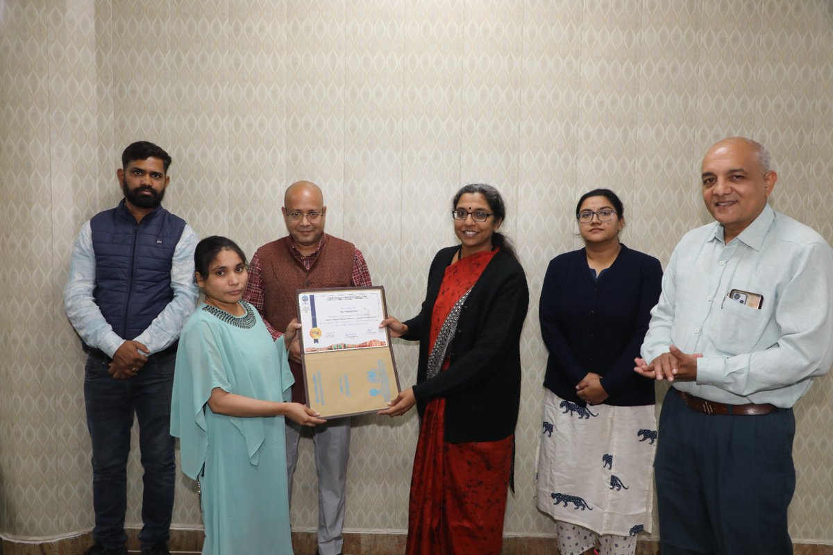
This initiative reflects CSIR-CDRI’s commitment towards capacity building and empowering young scientists in advanced life science research. @CSIR_IND @CsirSkill @IndiaDST @MSDESkillIndia #SkillDevelopment #ElectronMicroscopy #LifeSciences #TrainingProgram #CapacityBuilding
🔬 Ever wondered what battery materials look like on the inside? Cross sectioning reveals the secrets that surface analysis misses - unlocking better performance and safety. See how it works: azom.com/article.aspx?A… #BatteryResearch #ElectronMicroscopy
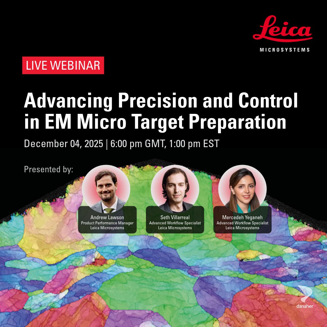
EM sample prep matters. Join our webinar to learn practical approaches and see how micro target preparation can improve precision and control. Register now: fcld.ly/px68j2w #ElectronMicroscopy #SamplePreparation #MicroTargeting

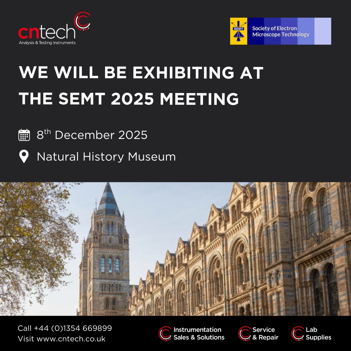
We are looking forward to exhibiting at the SEMT 2025 meeting and highlighting our electron microscopy solutions. The meeting will take place on the 8th of December at the Natural History Museum in London. Register here > rms.org.uk/rms-event-cale… #SEM #electronmicroscopy
Happy National Cake Day ! 🎂🍰🍓 (November 26) #ElectronMicroscope #electronmicroscopy Cryo-SEM of air bubbles and fat globules in fresh cream

We want to acknowledge the amazing #OpenScience #ElectronMicroscopy datasets by @alleninstitute, @HHMIJanelia, and our other sources! This work is a collaboration between myself @UCPH_health, @amreenmughal1 and @VanshikaChaddha @NIH, and Carolyn and Jennifer @HHMIJanelia.
Happy and honored to give a lecture at the 30th Congress of the Brazilian Society for Microscopy and Microanalysis in beautiful Maceió! #electronmicroscopy #microscopy #WomenInScience


Just a single, happy #eosinophil patrolling the human intestine! #electronmicroscopy #MicroscopyMonday #sciart
Here's a high-resolution peek at #neutrophils isolated straight from the human bloodstream, captured in stunning detail with #electronmicroscopy and colorized! 🤩 #MicroscopyMonday
Myelin sheath is a multilamellar spiral of cell membrane around an axon, consisting of alternating major dense and two intraperiod lines. Several diseases and conditions, such as multiple sclerosis, degrade myelin compromising the optimal electrical signal. #electronmicroscopy
Like a secret tucked into the bloodstream, #eosinophils (yellow) glow with singular beauty! #MicroscopyMonday #ElectronMicroscopy #sciart @EosinophilSoc

When the sentinel wakes! 🤩 An activated #neutrophil captured under #electronmicroscopy showing a uropod! #MicroscopyMonday #sciart
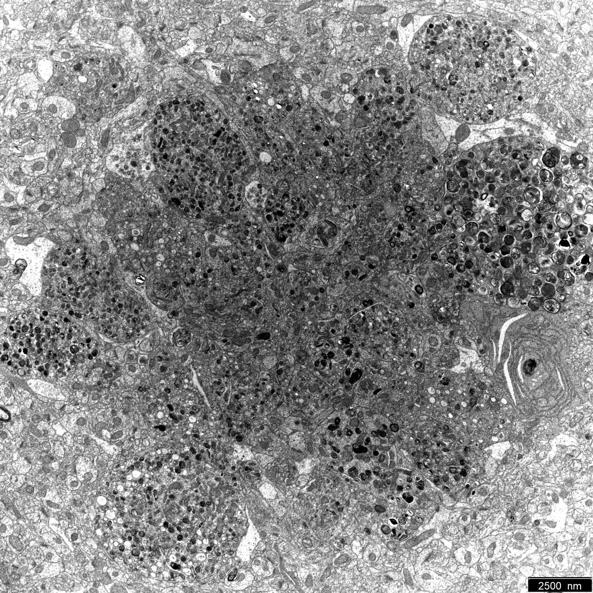
A high-res look inside an inflamed nasal tissue of a patient with chronic eosinophilic #rhinosinusitis! Disrupted #eosinophils and beautiful Charcot-Leyden crystals (blue) revealed by #ElectronMicroscopy! #Pathology #MicroscopyMonday


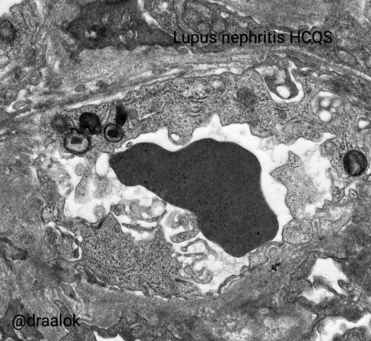
Identify this lesion in a patient of lupus nephritis #renalpath #electronmicroscopy @IndianRenal @Renalpathsoc @ISNkidneycare @Path_AIIMSDelhi

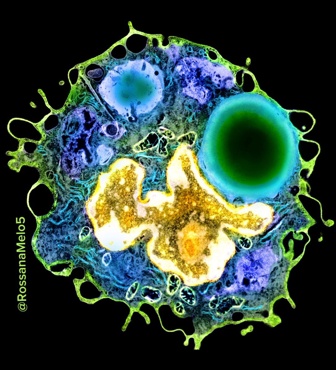
Starting the week strong with this powerful #macrophage full of phagosomes! 💪 #MicroscopyMonday #electronmicroscopy #bioart

If you are under 30 and interested in the nervous system, #myelin and #electronmicroscopy, we have three positions available at @Myelin_Alicante in @isabial_iis. 2-year contract near the beach! 🏝️ (and competitive salaries! 😱) #CLEM #volumeEM #AI #machinelearning #deeplearning


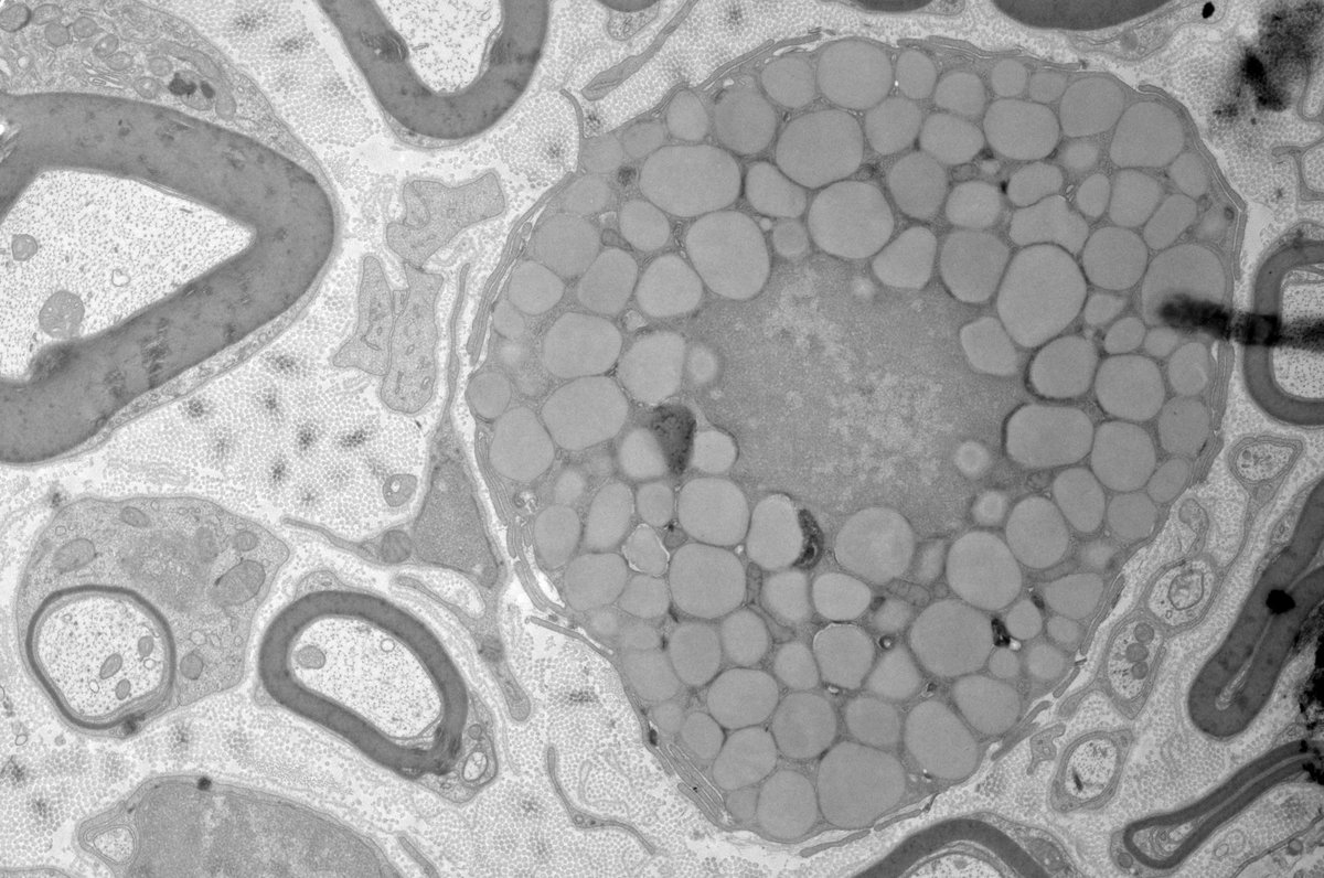
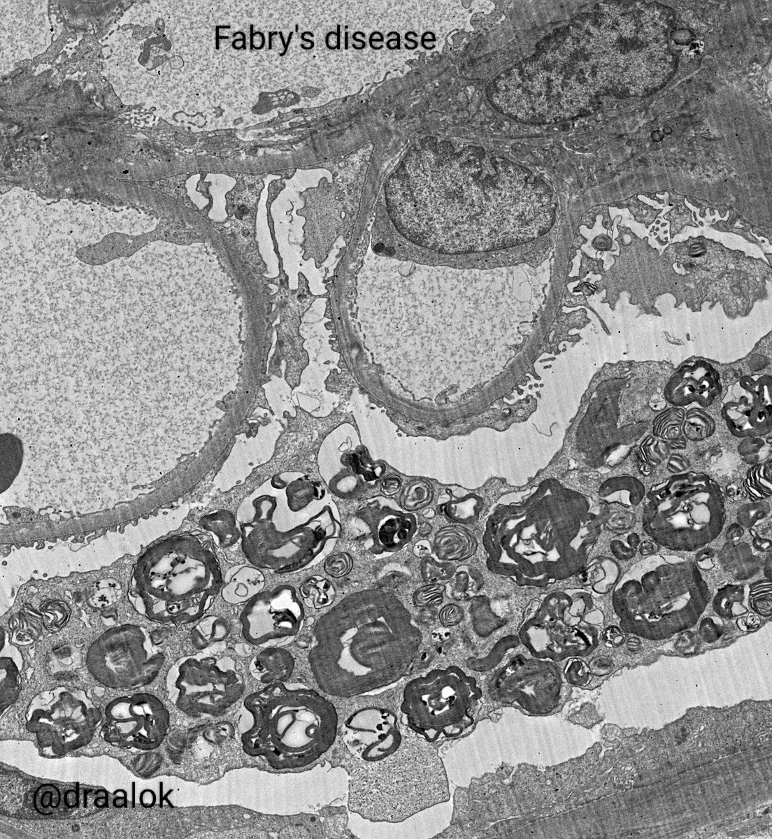
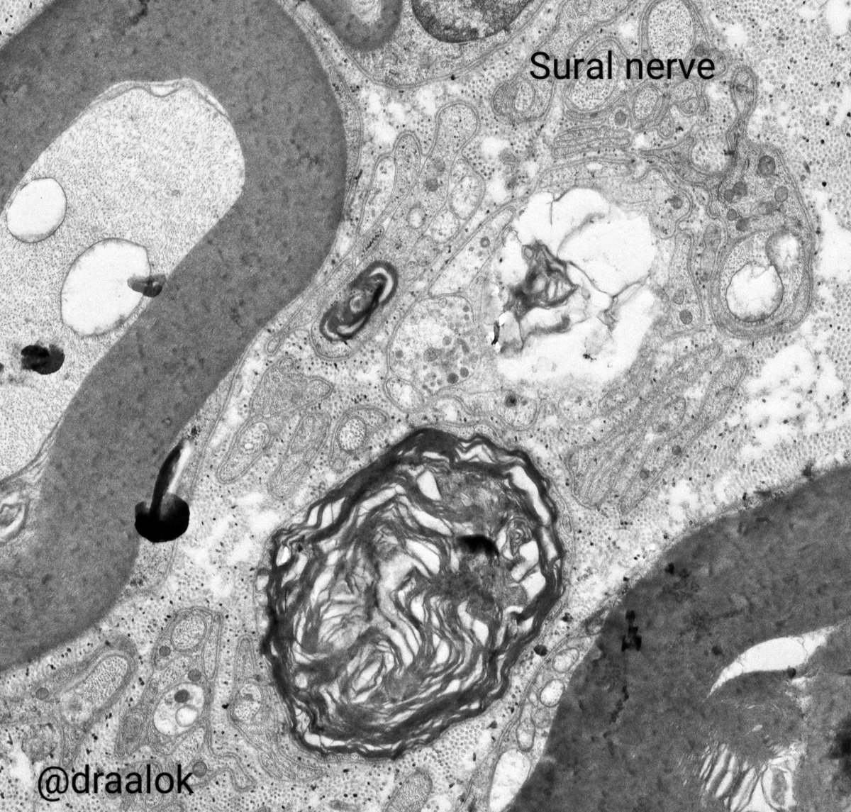
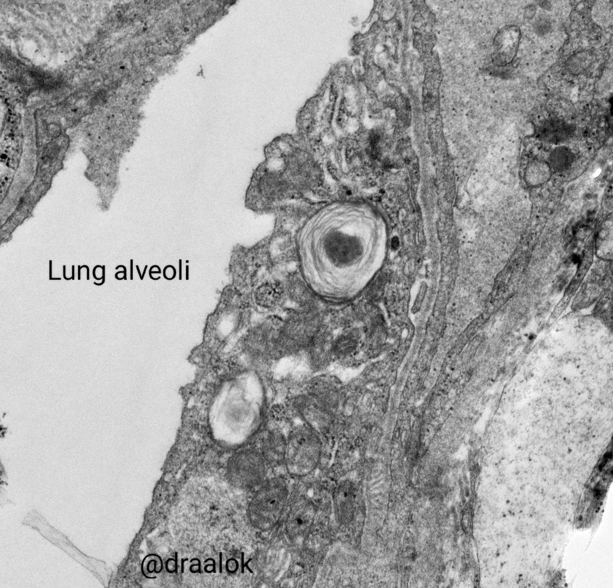
All in a days work! Diverse conditions featuring lamellated lysosomal inclusions. Kidney (Fabry's disease & HCQS toxicity in SLE/ lupus nephritis), Nerve (Amiodarone toxicity) and lung surfactant (normal) in alveoli! #nephtwitter #PathTwitter #electronmicroscopy #zebrabody
Amid the countless, one stands apart—, unique, radiant, and impossible to overlook. 🤩 #MicroscopyMonday #electronmicroscopy #eosinophil

My in-person talk at the @GordonConf vEM meeting is right around the corner! I will present @GSegala's, @LoogerL's, and my work: new chemically resistant fluorescent proteins (& enzymes!) for #CLEM / #ElectronMicroscopy. Come say hi! #Eos5 #hfYFP (and more) at #GRC #Microscopy

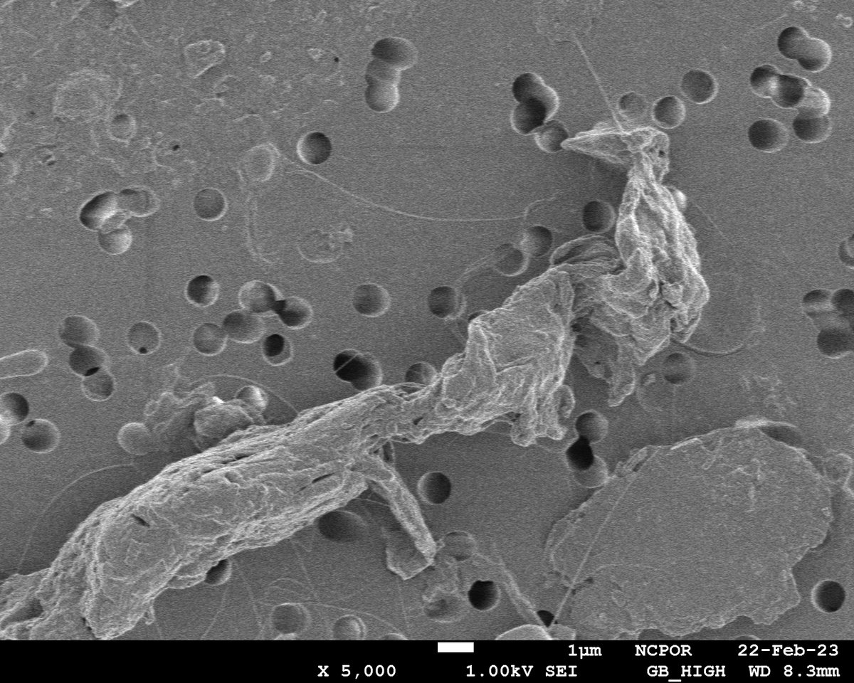
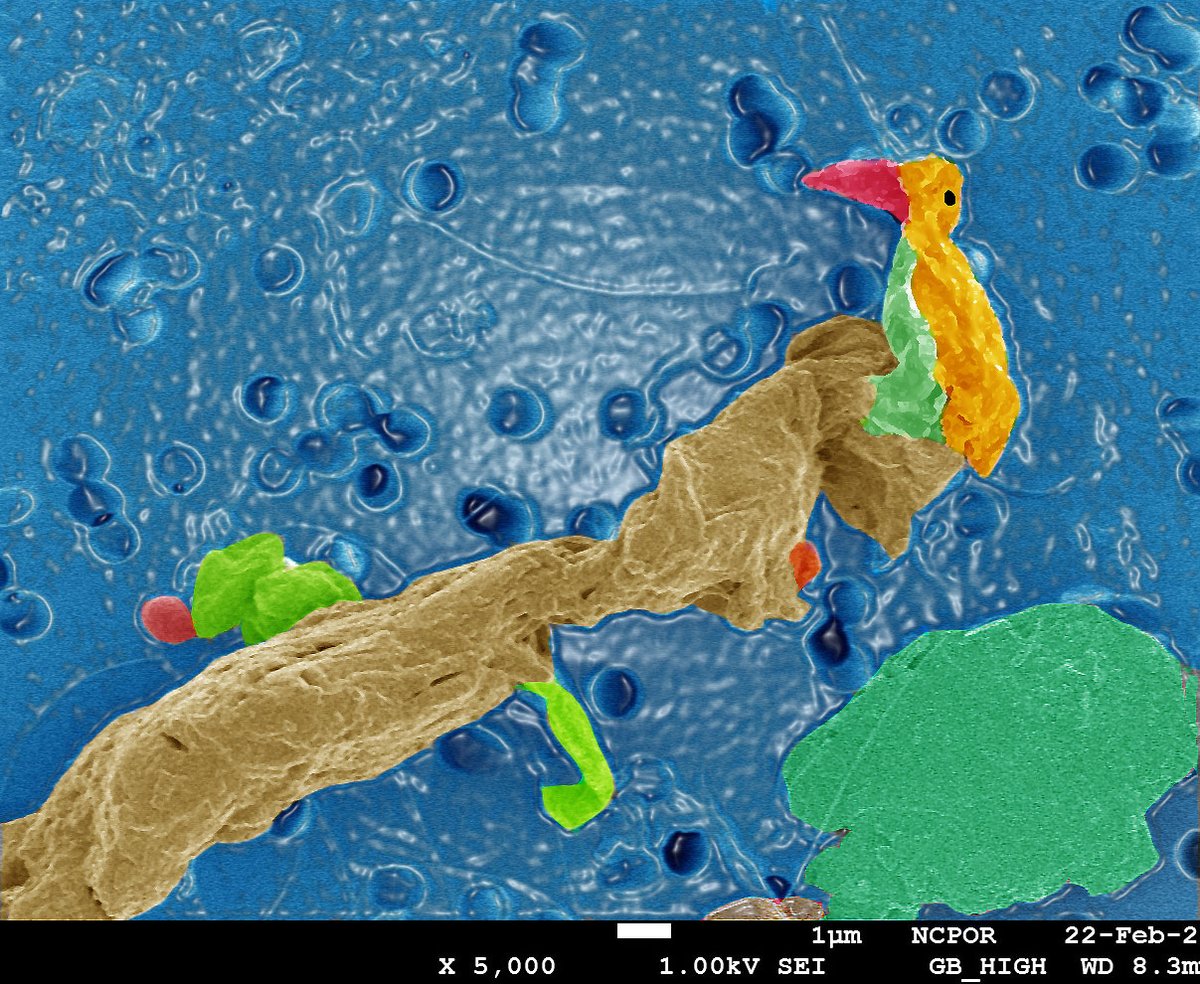
Filtrate on filter paper is a key step to prepare water sample for #electronmicroscopy analysis. However, interpretation takes on a new meaning such as this colored art (a perching bird enjoying pond view on a rainy day) made from a filter paper residue. Colors do create emotion.

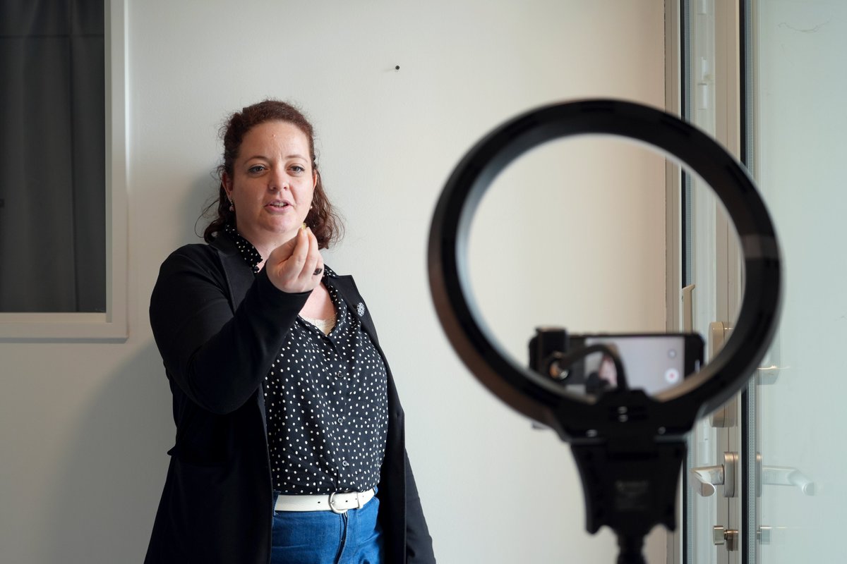
📹Rolling.... and Action! Imaging scientists are still on the set for our workshop "FBI training" about video producing. Here, @MelinaPetrel explains ultramicrotomy for a future MOOC on #electronmicroscopy🔬
Starting the week with these nice membranes is one of the best things it could have happened to me. Finally happy 🥰 #ElectronMicroscopy
Melainia McClain, a #scientist in our #ElectronMicroscopy team, advises & assists our researchers with a wide range of 3D EM imaging techniques. #microscopy #SerialSectioning #innovation #ArrayTomography #SPOTLIGHTSTOWERS
Happy National Cake Day ! 🎂🍰🍓 (November 26) #ElectronMicroscope #electronmicroscopy Cryo-SEM of air bubbles and fat globules in fresh cream
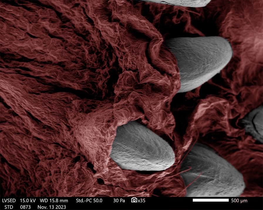
It's #MicroscopyMonday again 🔬 Today we're sharing this image of a strawberry, in red false colour, with strawberry seeds 🍓 taken on our @JEOLEUROPE IT510 LV SEM! #ElectronMicroscopy #Strawberry #Food #Biology

✨Ready for our next #Brainingproject seminar? 🧠 We are excited to welcome Dr. @ConchiLillo from @Incyl_Usal Salamanca,Spain. Dr. Lillo is expert in #electronmicroscopy and retinal pathologies. Join us tomorrow 👇🏽 📅Thursday, 21/11 🕕18h CET | 17h Lisbon time #cofundedbyEU


There’s fascinating battery research being presented at the UK’s Faraday Institution Conference. Our specialists appreciate discussing how we can help materials analysis challenges with Electron Microscopy, NMR, Raman and AFM innovations #NMRchat #ElectronMicroscopy #Raman #AFM

Something went wrong.
Something went wrong.
United States Trends
- 1. Broncos 45.8K posts
- 2. Mariota 12.8K posts
- 3. Ertz 3,073 posts
- 4. Commanders 32.7K posts
- 5. Bo Nix 9,834 posts
- 6. #RaiseHail 5,697 posts
- 7. Riley Moss 2,262 posts
- 8. #BaddiesUSA 24.6K posts
- 9. Bobby Wagner 1,043 posts
- 10. Burks 15.2K posts
- 11. Terry 20.3K posts
- 12. #DENvsWAS 3,165 posts
- 13. Deebo 3,089 posts
- 14. Bonitto 5,580 posts
- 15. Collinsworth 3,016 posts
- 16. #RHOP 12K posts
- 17. Sean Payton 1,608 posts
- 18. Happy New Month 144K posts
- 19. Dan Quinn N/A
- 20. Zach Edey 3,001 posts