#microscopy search results

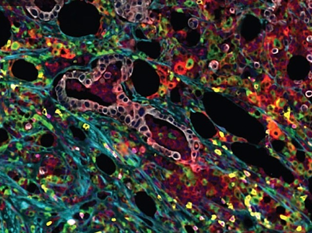
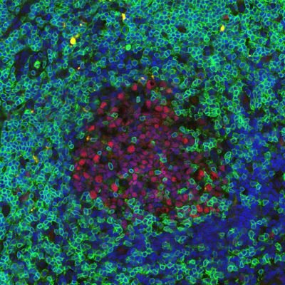
Can solid tumors become CAR T therapy targets? Learn how researchers identified the uPAR molecule in solid tumors with spatial precision. Image using Leica Microsystems Cell DIVE multiplex imaging. 🔗fcld.ly/og8dkii @BPoD_s @BiochemOxford #Microscopy #LifeScience

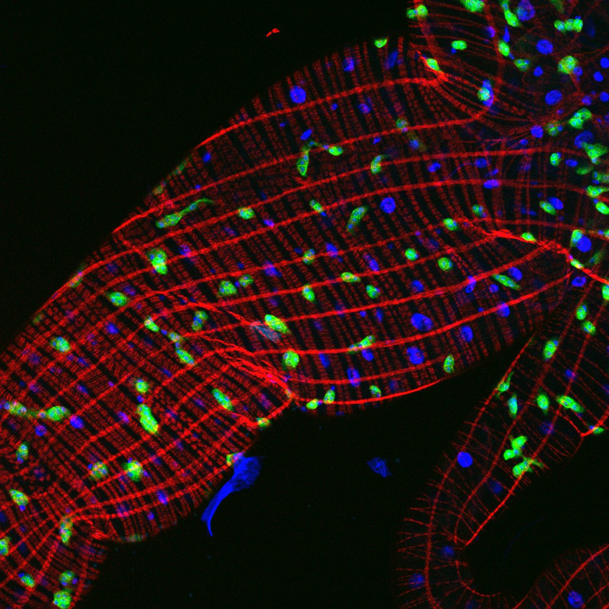
Immuno-Fl. (Cardiac M-line by Titin) | Rat cardiomyocyte ・Titin (Proteintech, 27867-1-AP) + AF488: Green ・Phalloidin 633: Magenta ・Hoechst: Blue by BZ-XLC1, confocal #PositiveControlSharing #Microscopy #Titin #ポジコン共有 #顕微鏡


Boutin, Kodjabachian et al. describe an inducible multiciliated cell line well suited for advanced #microscopy and #proteomic approaches. The study provides a detailed proteomic profiling of MCC during their differentiation. hubs.la/Q043ClPD0 #Cilia


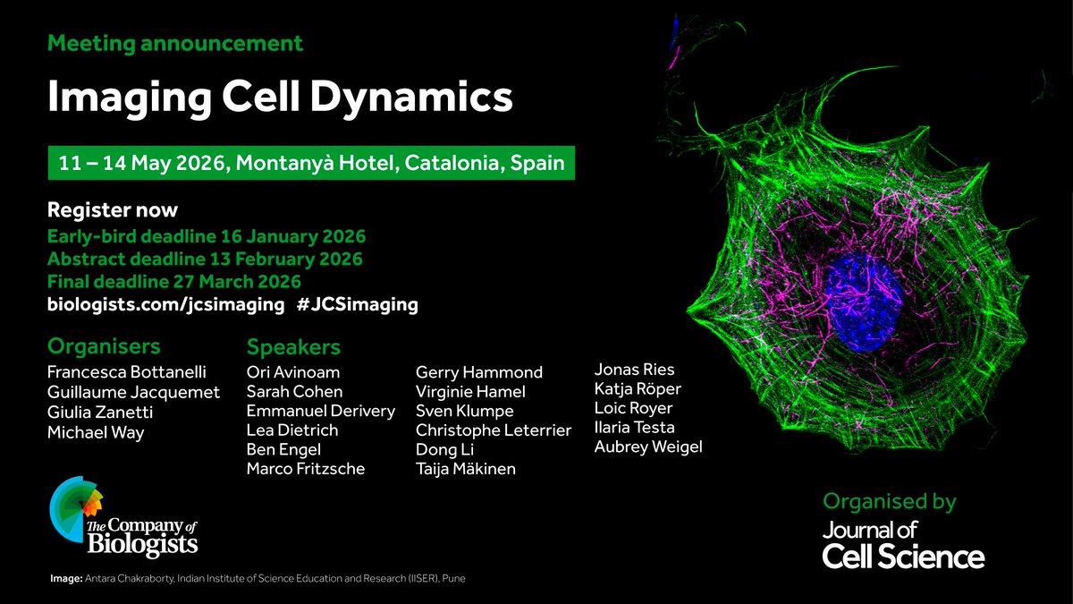
The abstract submission deadline for our upcoming 'Imaging Cell Dynamics' meeting is this Friday. We still have a few places remaining so apply now to secure your spot! Find out more at: biologists.com/meetings/jcsim… #imaging #microscopy #cellbiology

#AudioVisualAbstract : Incident Dark-Field (IDF) Illumination #Microscopy as a Novel Tool for Real-Time, Dye-Free #ParathyroidPerfusion Assessment in #ThyroidSurgery rdcu.be/feFzU @TraceyPuMD @SyedAAhmad5
Interplanetary Marauders and other demos of the Turing complete nature of the PCS (PARDUS Command Script) programming language feature in my latest YouTube short: youtube.com/shorts/jZY9kpi… #Coding #Microscopy


An Editors' Pick via #OPG_OpEx: High-fidelity super-resolution optical fluctuation imaging via frequency separation correlation bit.ly/4qx5lPd #SuperResolution #Microscopy.


#BPS2026 Newsroom Spotlight - New Microscopy Technique Lets Scientists See Cells in Unprecedented Detail & Color @debsankarsahaR1 @maxprigozhin @mcbHarvard #microscopy#CryoEM #BiologicalImaging buff.ly/es8fJEY


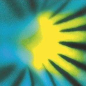
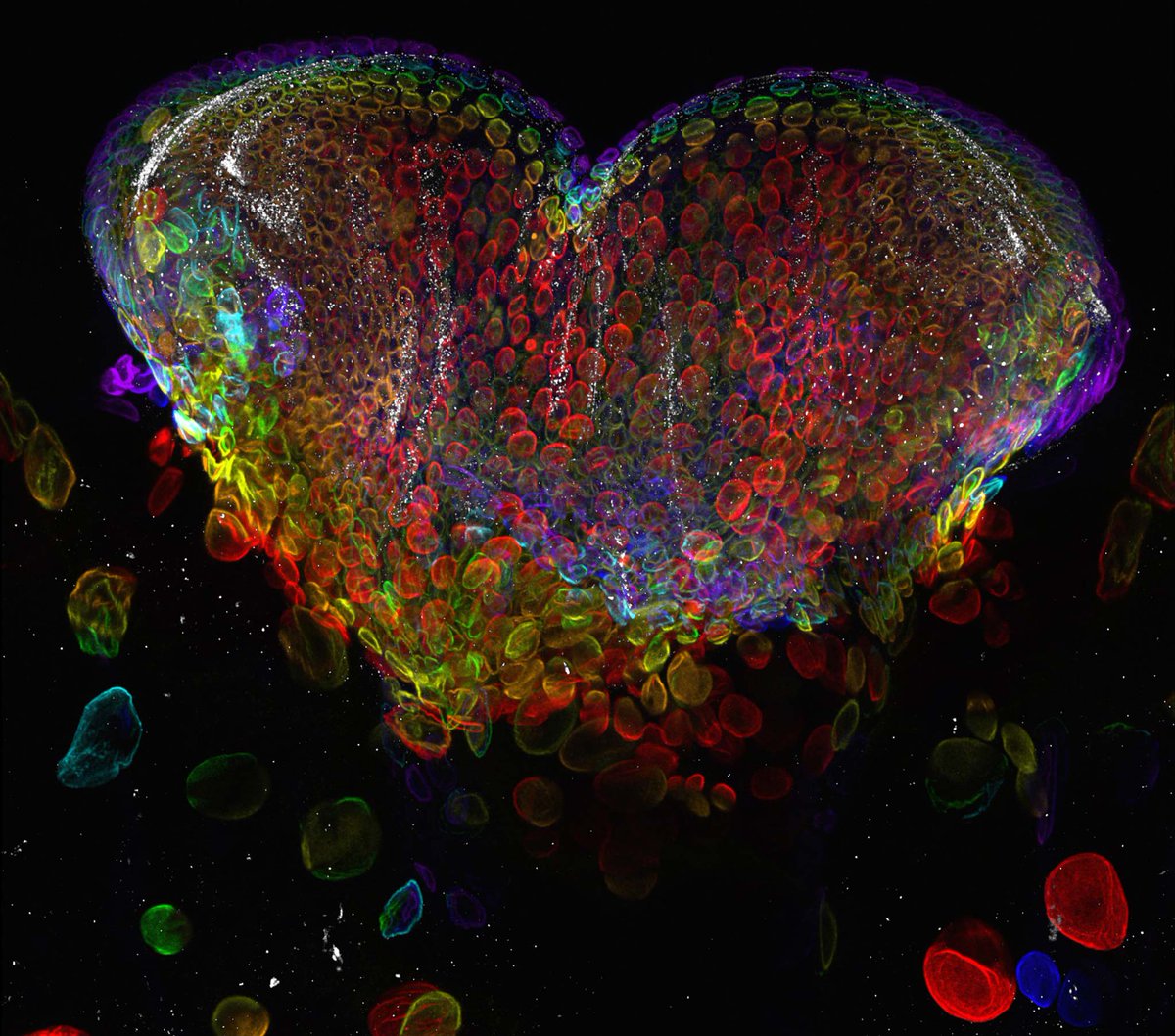
Guess you can use a timeline cleanser? Can I show you some microscopy? My job is to see beauty even in sad things (so we can learn how to prevent them!). This is a brain with early Alzheimer. This image was acquired in 3D and took 72 hours to get ready! #Microscopy #Neuroscience


Now You See Me Super-Resolution Microscopy Next-generation versions with increased capabilities and better workflow integration are hitting the market @bruker @massphoton @NikonInst @oniHQ #microscopy #AI hubs.li/Q03-2mwb0


Scientists have introduced a microscopy technique that reveals a hidden layer of chemistry involving molecules that normally evade detection. A team at the University of… dlvr.it/TS0LBN #Microscopy #MolecularScience #Biochemistry #TokyoUniversity #ResearchBreakthrough

How do researchers prepare complex 3D samples for cryoEM? Watch the on-demand webinar to see how high-pressure freezing, CLEM, and 3D targeting fit into practical workflows. Discover more: fcld.ly/wr196or #CryoEM #Microscopy


Sharing a bit of my journey and life in Wuhan. From the lab at the Institute of Hydrobiology @CAS__Science to walks along East Lake, it has been a rewarding experience balancing research and family life.🔬🌿 #ResearchLife #Wuhan #Microscopy #MayDay

Mexican Cesar Valades has settled in Wuhan after studying in France. By using AI & microscopy technologies, Cesar dedicates his expertise to study of aquatic ecosystems. Diligent work knows no borders; may all workers around the world have a joyful May Day holiday!

🔬 Acquisition in CA1 and ACC regions with over 200 cells in CA1 identified across multiple days, and ~80% of cells registered across multiple sessions. Acquired with Quartet. Courtesy of Gal Elyasaf, Ph.D., Weizmann Institute. #MicroscopyMonday #Neuroscience #Microscopy

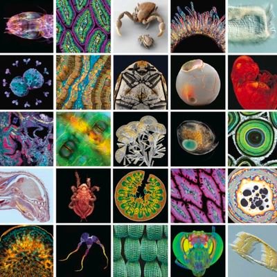
Step into the microscopic world 🌌 A hidden universe of beauty and wonder exists all around us - too small for the naked eye.Mind-blowing details everywhere! #MicroscopicWorld #Microscopy #Aiart #GrokImagine

Most of what we “see” in 3D #microscopy is wrong. But a new preprint from the lab driven by Anna Pittman comes to the rescue. Hard truth: humans are bad at seeing 3D on screens. We collapse volumes, look at image slices and pretend it’s enough. But, a model can interrogate the


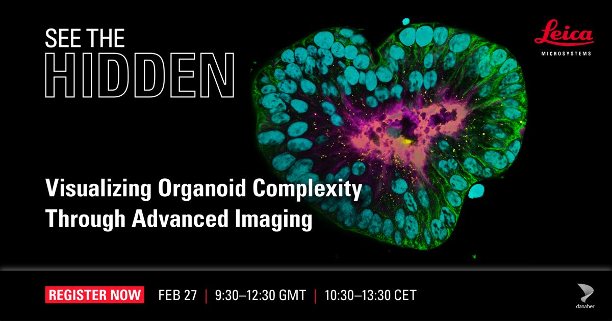
🚀 Spoiler alert: something new is coming! Join our See the Hidden hybrid event with @MicronOxford to explore new imaging tools for #Organoid research, including the next generation of #LightSheet #Microscopy 📅 Feb 27 | 09:30 GMT🔗 Register fcld.ly/5h6kf91

Our how-to for observing a meteorite through a microscope. dlvr.it/TRQtj0 #Meteorites #Stargazing #Microscopy #SpaceExploration #ScienceEducation


HAVE YOU SEEN? 4 NEW COURSES ADDED TO OPENLEARN! 🔬 Digital fluorescence #microscopy 🏃♀️ Extreme #endurance performance 💻 Using @Turnitin effectively (for #students) 🗨️ #Language in professional life All available via the 'New to OpenLearn' page 👇 open.edu/openlearn/get-…

Sub-nanometer imaging is pushing the limits of what we can see—down to molecular and atomic scales. Explore key techniques like AFM, EELS, and TERS transforming nanotech & biophotonics: findlight.net/blog/sub-nanom… #Photonics #Nanotechnology #Microscopy

Immuno-Fl. (Cardiac M-line by Titin) | Rat cardiomyocyte ・Titin (Proteintech, 27867-1-AP) + AF488: Green ・Phalloidin 633: Magenta ・Hoechst: Blue by BZ-XLC1, confocal #PositiveControlSharing #Microscopy #Titin #ポジコン共有 #顕微鏡
Interplanetary Marauders and other demos of the Turing complete nature of the PCS (PARDUS Command Script) programming language feature in my latest YouTube short: youtube.com/shorts/jZY9kpi… #Coding #Microscopy
Can solid tumors become CAR T therapy targets? Learn how researchers identified the uPAR molecule in solid tumors with spatial precision. Image using Leica Microsystems Cell DIVE multiplex imaging. 🔗fcld.ly/og8dkii @BPoD_s @BiochemOxford #Microscopy #LifeScience
Sharing a bit of my journey and life in Wuhan. From the lab at the Institute of Hydrobiology @CAS__Science to walks along East Lake, it has been a rewarding experience balancing research and family life.🔬🌿 #ResearchLife #Wuhan #Microscopy #MayDay
Mexican Cesar Valades has settled in Wuhan after studying in France. By using AI & microscopy technologies, Cesar dedicates his expertise to study of aquatic ecosystems. Diligent work knows no borders; may all workers around the world have a joyful May Day holiday!

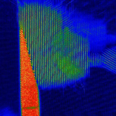
This result deserves sharing on #fluorescencefriday. I bet you can notice brighter fluorescent signal with an ultrafast laser that was optimized for a special brain imaging application and a microscope. There will be more details. Stay tuned! #microscopy #neuroscience #2p 💕

DYNASTY creates hands-on learning opportunities in microscopy and data analysis, turning training into practical experience in the lab. #DYNASTYProject #HandsOnTraining #Microscopy #2Dmaterial #HorizonEurope

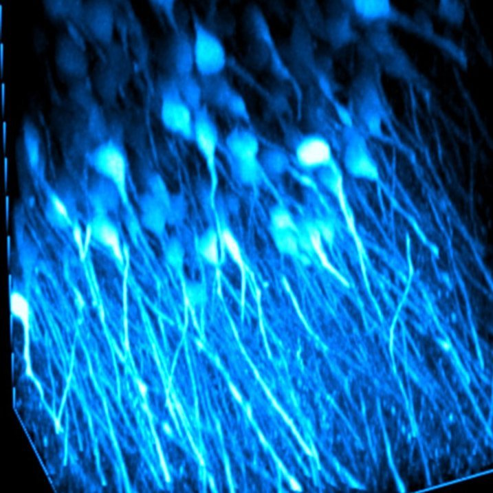
🖥️ 𝗜𝗹𝗹𝘂𝗺𝗶𝗻𝗮𝘁𝗶𝗻𝗴 𝗕𝗿𝗮𝗶𝗻 𝗙𝘂𝗻𝗰𝘁𝗶𝗼𝗻 𝗧𝗵𝗿𝗼𝘂𝗴𝗵 𝗧𝘄𝗼-𝗣𝗵𝗼𝘁𝗼𝗻 𝗠𝗶𝗰𝗿𝗼𝘀𝗰𝗼𝗽𝘆: 𝗖𝗮𝗹𝗰𝗶𝘂𝗺 𝗮𝗻𝗱 𝗩𝗼𝗹𝘁𝗮𝗴𝗲 𝗗𝘆𝗻𝗮𝗺𝗶𝗰𝘀 𝘄𝗶𝘁𝗵 𝗢𝗽𝘁𝗼𝗴𝗲𝗻𝗲𝘁𝗶𝗰𝘀 ▶️ goto.bruker.com/4nmSTBf #neuroscience #cellbiology #microscopy

Our latest preprint list is now up on FocalPlane. This week, bioimage analysis takes centre stage! focalplane.biologists.com/2026/05/01/mic… #microscopy #bioimageanalysis #preprints


Our latest issue of infocus Magazine #RMSinfocus is now freely available to read online - featuring a range of the most impactful outreach and education initiatives within the RMS and wider #microscopy community. Read more: ow.ly/XlNw50YT9Sr

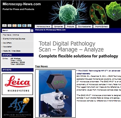
🔬 The #digitalpathology & AI-assisted #microscopy market is accelerating with smarter #diagnostics, automated slide analysis, and AI-powered clinical precision. Digital diagnostics is shaping the future of #pathology. knowledge-sourcing.com/report/digital… #Healthcare #MarketInsights #Medical


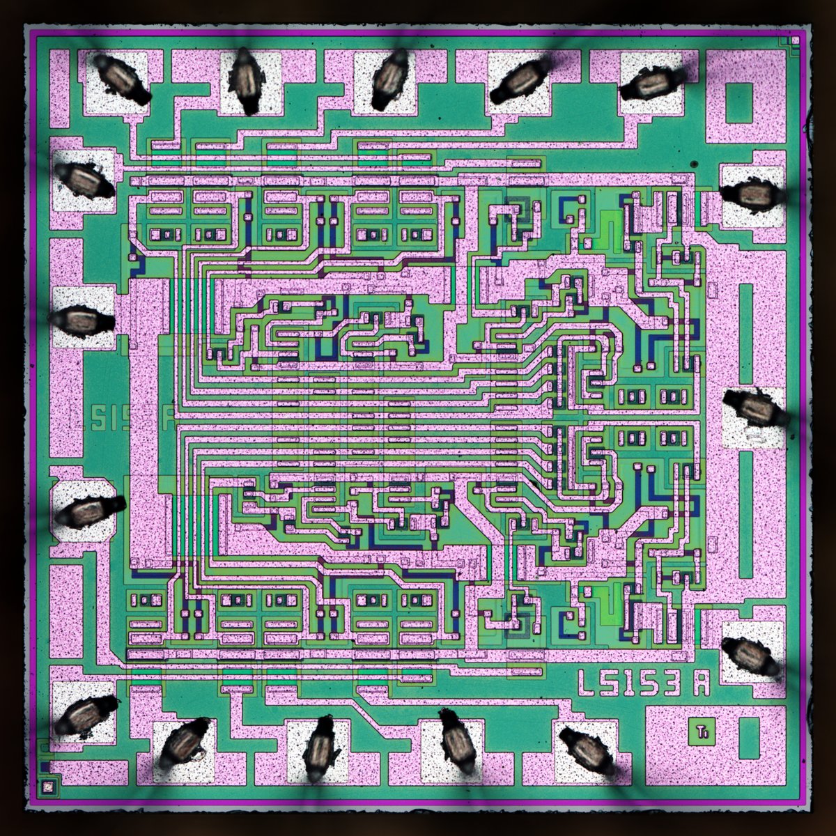
Ti 54LS153 - Dual 4→1 Data Selectors/Multiplexers : weekend die-shot zeptobars.com/en/read/Ti-54L… via @Zeptobars @TXInstruments #electronics #microscopy

🔬 Dive deep into female anatomy! Vaginal secretions are complex biological ecosystems, revealing a surprising mix of cells, bacteria, and natural pH balance under the microscope. Nature's chemistry is wild. #ScienceFacts #Biology #Microscopy #HumanBody

Crafting Excellence. Honoring Dedication. Happy Labor Day from ModuleSci #ModuleSci #Microscopy #TechLife #MayDay #HumanDetail #Innovation


Penicillium Slide Identification Microbiology practicals ka important topic! Is post mein Penicillium ke key microscopic features simple way mein explain kiye hain for quick revisions & better identification. #Microbiology #Penicillium #Microscopy #StudyThreads #MedicalEducation

🚨 Deadline Extended! 🚨 The Cilia 2027 Best Image Competition is now open until May 7, 2026. Got a striking microscopy image? This is your chance to have it showcased at #Cilia2027 🦠🏆 Submit now and share your science through art! #Microscopy #SciArt #CellBiology #imaging


🎙️ #IBIWebinarSeries Join us for an exciting talk by Dr. @BuvanSR (Northwestern University) 🧠 Vimentin IFs in motion—uncovering cytoskeletal dynamics 📅 15 May 2026 | ⏰ 3–4 PM IST 🔗 Register (Free): indiabioimaging.org/upcoming-events #IndiaBioImaging #Microscopy #Bioimaging


My image, "Cellular Cosmic Body," has received an Honorable Mention in the Evident Image of the Year Award. Highlighting radiating Actin and delicate Septin filaments Want your research to have a similar treatment? DM me to collaborate! #SciArt #Microscopy #EvidentIOTY

Microbiology practicals ka important topic Is post/reel mein Plasmodium falciparum ke microscopic identification points simple way mein explain kiye hain for quick revision and viva prep #Microbiology #PlasmodiumFalciparum #Microscopy #MedicalEducation #StudyThreads

📢 #LAQV Seminars | May 5, 14h WET 🔬 Join us! 👉 Benito Rodríguez on Electron #Microscopy (SEM, TEM, FIB) for materials #characterization 👩💻 Chairs: J. F. Lodeiro & H. M. Santos 🏛️ NOVA FCT, Room 217 D 🔗 laqv.requimte.pt ✨ #ScienceForFuture #Innovation

Disclosure and the Hybrid Constitution of UK Company Law gsdiandadvocacy.co.uk/from-property-… #microscopy #sciencefacts #biologyteacher #physics #Fitness #Gym #Workout #Fitsporation #Bodybuilding #FitnessMotivation #Yoga #GymLife #CrossFit #FitnessModel #FitnessAddict #PersonalTrainer


Immuno-Fl. (Cardiac M-line by Titin) | Rat cardiomyocyte ・Titin (Proteintech, 27867-1-AP) + AF488: Green ・Phalloidin 633: Magenta ・Hoechst: Blue by BZ-XLC1, confocal #PositiveControlSharing #Microscopy #Titin #ポジコン共有 #顕微鏡
Can solid tumors become CAR T therapy targets? Learn how researchers identified the uPAR molecule in solid tumors with spatial precision. Image using Leica Microsystems Cell DIVE multiplex imaging. 🔗fcld.ly/og8dkii @BPoD_s @BiochemOxford #Microscopy #LifeScience
Boutin, Kodjabachian et al. describe an inducible multiciliated cell line well suited for advanced #microscopy and #proteomic approaches. The study provides a detailed proteomic profiling of MCC during their differentiation. hubs.la/Q043ClPD0 #Cilia
On the cover of our #LatestIssue: Fluorescent confocal #microscopy showing spatial expression patterns of AtSWEET1 in an embryo isolated from a germinating #Arabidopsis thaliana seed. Image courtesy of Xueyi Xue. 📖 See Chen et al. nph.onlinelibrary.wiley.com/doi/10.1111/np…

#LatestIssue cover: #Microscopy image showing DR5:3nucVenus expression in the inflorescence apical meristem of an Arabidopsis thaliana plt3 plt5 plt7 pin1-T600I mutant. Image courtesy of Kerstens & Willemsen. 📖 See @MHL_Kerstens & @ViolaWillemsen et al. doi.org/10.1111/nph.70…

The abstract submission deadline for our upcoming 'Imaging Cell Dynamics' meeting is this Friday. We still have a few places remaining so apply now to secure your spot! Find out more at: biologists.com/meetings/jcsim… #imaging #microscopy #cellbiology

✨A gut feeling you’ll love this one 🐛 The anterior midgut of a Drosophila melanogaster 🪰larva puts on a confocal glow show! 🟢 Adult midgut progenitors 🔴 Actin 🔵 Nuclei 📸Image by Sapna Krishnakumar #FluorescenceFriday #Microscopy #DevBio
Would your #cellbio research benefit from you attending a #Microscopy or #BioimageAnalysis training course? For grants of up to £1,000, check out our Microscopy Training Grants with @focalplane_jcs. Next deadline: 5 September 2025 biologists.com/grants/jcs-foc…


In this My Research Using Nikon's Microscopes, W. Gregory Sawyer, PhD (Moffitt Cancer Center) shares his perspective ✨ Read more ▶ bit.ly/4rn0YH7 *Position and affiliation mentioned are as of the website publication time. #NikonMicroscopy #Microscopy

Registration for our Imaging Cell Dynamics meeting is still open! Organised by Francesca Bottanelli, Guillaume Jacquemet, Michael Way and Giulia Zanetti. Find out more: biologists.com/meetings/jcsim… Early-bird deadline: 16 January #imaging #microscopy

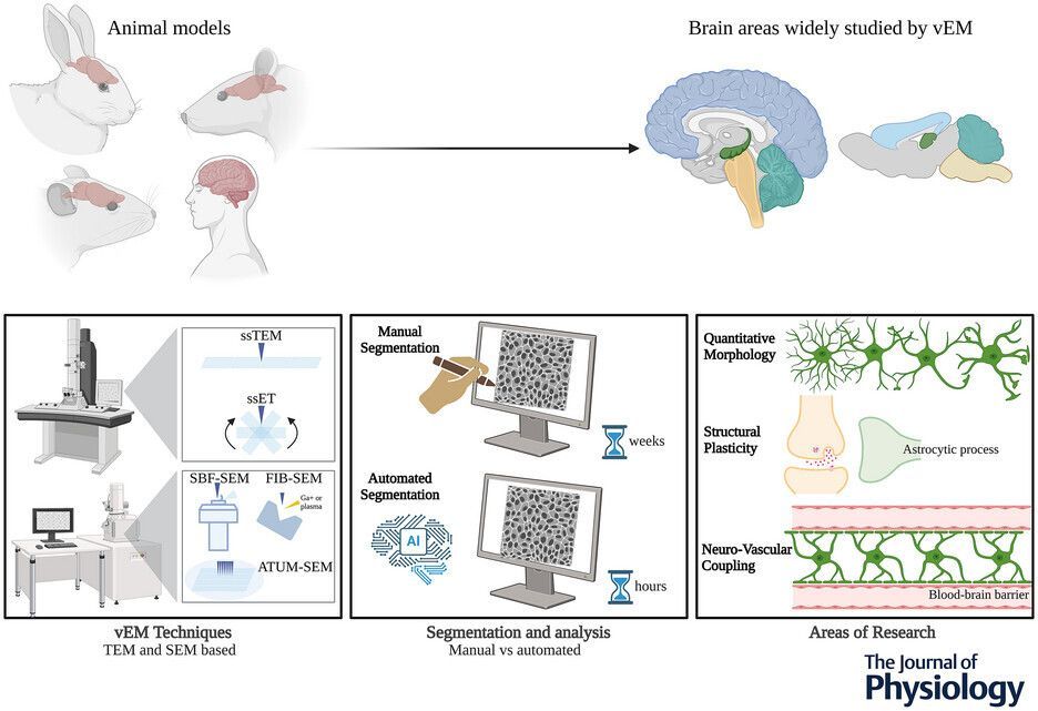
Vanessa Chiappini (@unito) and co-authors present this scoping review into ultrastructure of #astrocytes using volume electron #microscopy 📜 🔬 🔗 Read the paper: buff.ly/3KvTqP4
An Editors' Pick via #OPG_OpEx: High-fidelity super-resolution optical fluctuation imaging via frequency separation correlation bit.ly/4qx5lPd #SuperResolution #Microscopy.

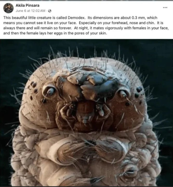
You are not alone. 😨🛑 Meet Demodex, the microscopic creatures living in your pores right now. They mate, lay eggs, and die on your face. #Microscopy #Biology #HiddenWorld
Guess you can use a timeline cleanser? Can I show you some microscopy? My job is to see beauty even in sad things (so we can learn how to prevent them!). This is a brain with early Alzheimer. This image was acquired in 3D and took 72 hours to get ready! #Microscopy #Neuroscience
Sub-nanometer imaging is pushing the limits of what we can see—down to molecular and atomic scales. Explore key techniques like AFM, EELS, and TERS transforming nanotech & biophotonics: findlight.net/blog/sub-nanom… #Photonics #Nanotechnology #Microscopy

L'œil d'une mouche Drosophila melanogaster au 3e stade larvaire, en #microscopie confoncale. Joyeuse Sain-Valentin ! Crédits : Dr. Michael John Bridge nikonsmallworld.com/galleries/2012… #microscope #microscopy #insectes #insects #entomology #entomologie
Great to be here in Reading for Dynamic Cell VI, organised jointly by our friends at the Biochemical Society and the British Society for Cell Biology (BSCB) 🙂 Come and chat with us about the benefits of RMS membership and our range of activities in support of #microscopy 🔬


🐖Structural and Functional Characterization of Porcine Adeno-Associated Viruses by Nelson et al. ✍️mdpi.com/1999-4915/17/9… #porcine #virus #microscopy #gene

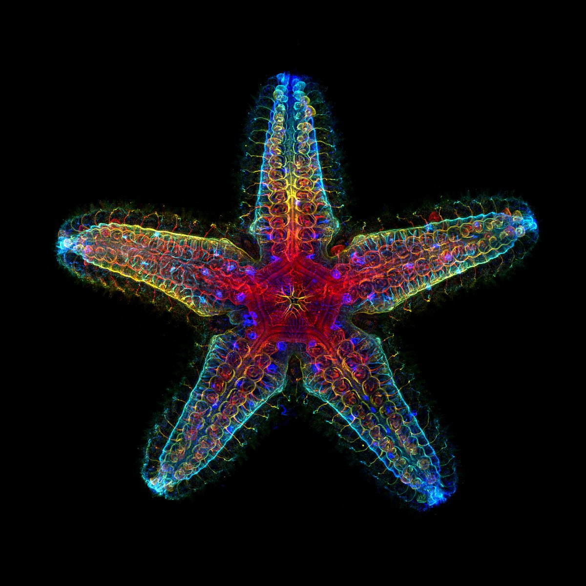
Le système nerveux d'une jeune étoile de mer, visualisé en microscopie confocale à fluorescence ⭐🔬 Crédits : Laurent Formery & Nathaniel Clarke via @nikonsmallworld nikonsmallworld.com/galleries/2024… #microscope #microscopy #nature #artandscience #beautifulscience
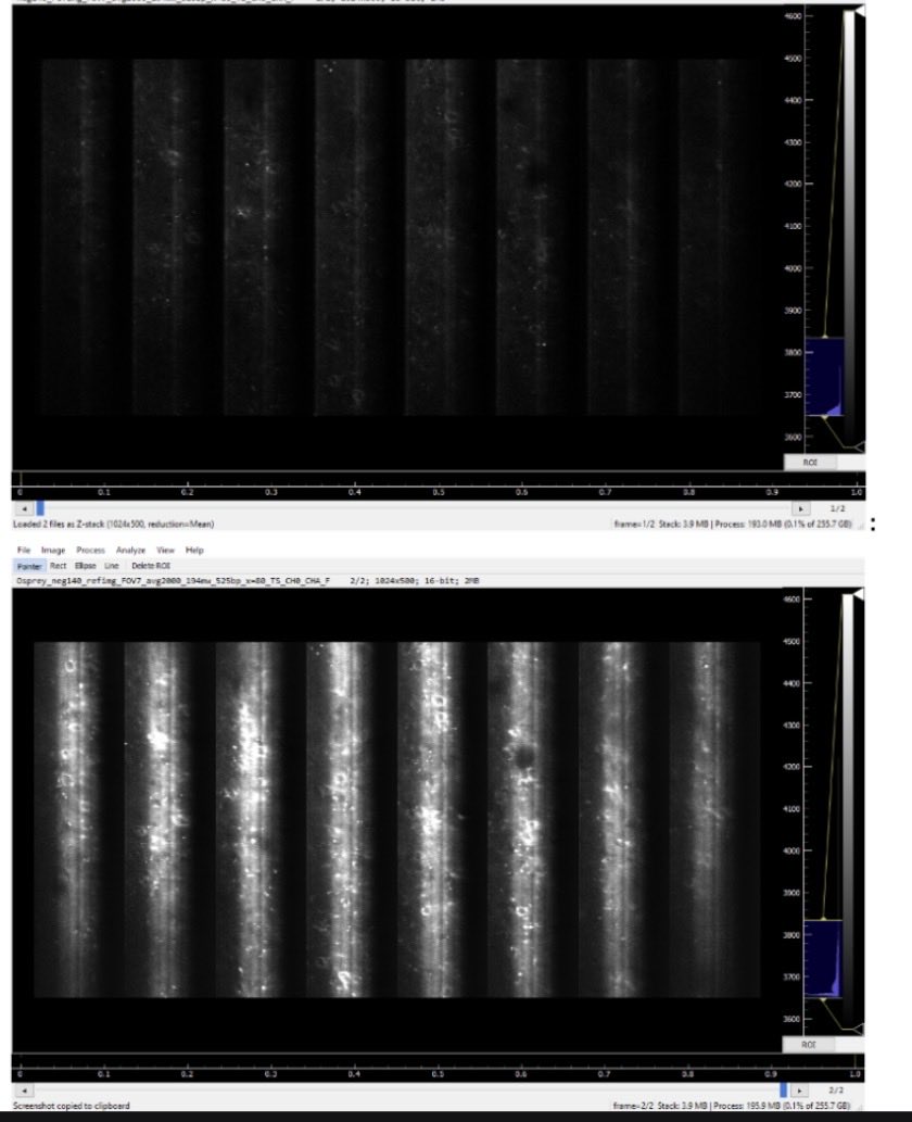
Seeing photoresist residue on wafers can be difficult and negatively impact inspection. For efficient visualization of residual contamination, fluorescence #microscopy has advantages over conventional imaging methods. Discover more: fcld.ly/tq20sr8 #WaferInspection

Something went wrong.
Something went wrong.
United States Trends
- 1. #euphoria N/A
- 2. #GoAvsGo N/A
- 3. #GoAvsGo N/A
- 4. BEST OF ME N/A
- 5. Jarrett Allen N/A
- 6. #mnwild N/A
- 7. Rudy N/A
- 8. May the 4th N/A
- 9. #RHOA N/A
- 10. Cale Makar N/A
- 11. Jules N/A
- 12. Edmond N/A
- 13. Wallstedt N/A
- 14. Quinn Hughes N/A
- 15. Wally N/A
- 16. Raptors N/A
- 17. Lou Ann N/A
- 18. Lexi N/A
- 19. Habs N/A
- 20. Vijay N/A