#imageofthemonth resultados de búsqueda

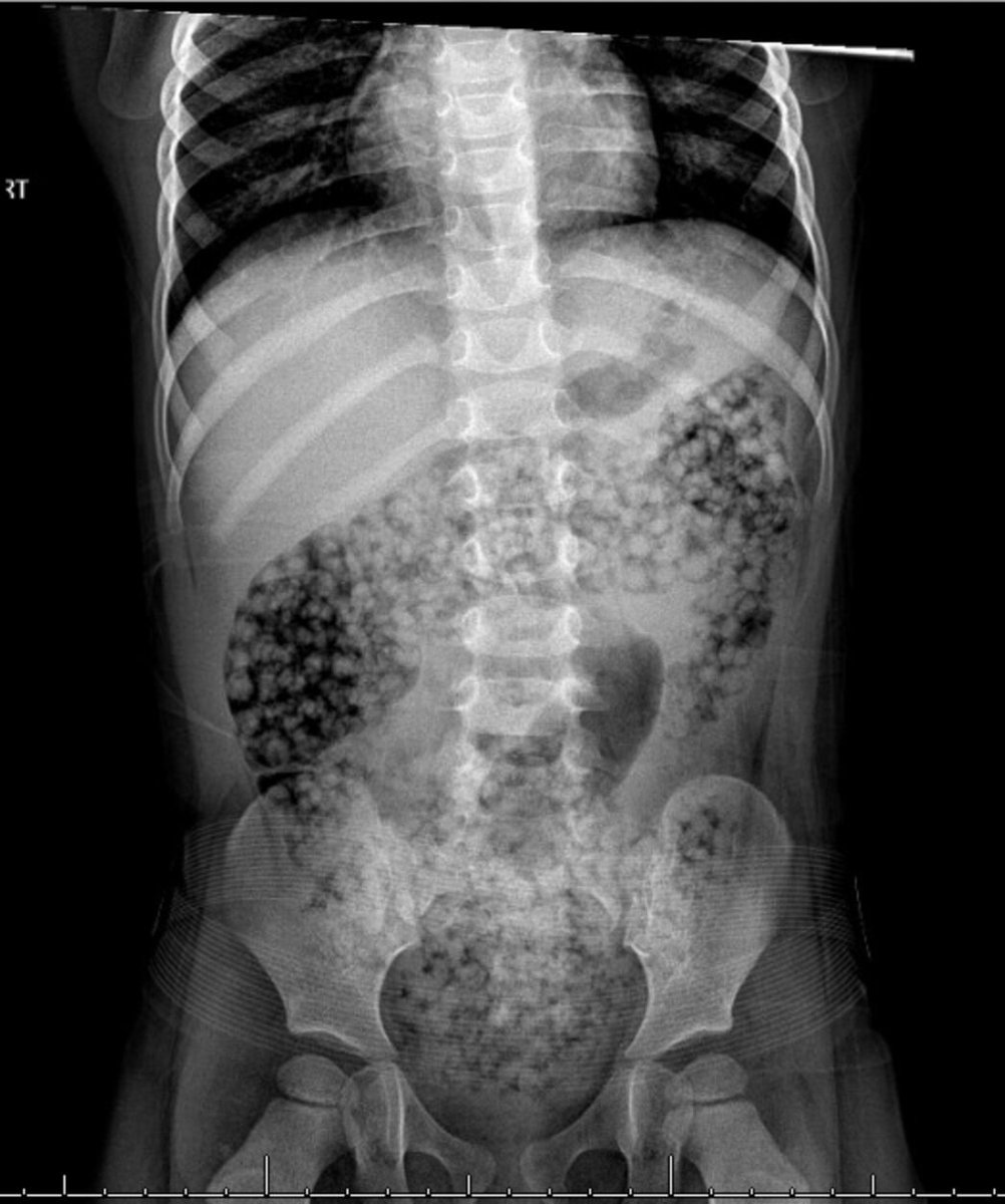
3yo girl ingested water beads. Now has abdominal pain, emesis. X-ray shows beads in large intestine. First-line management? Answer and learn more 👉 bit.ly/48ivJTe #imageofthemonth #quickpoll
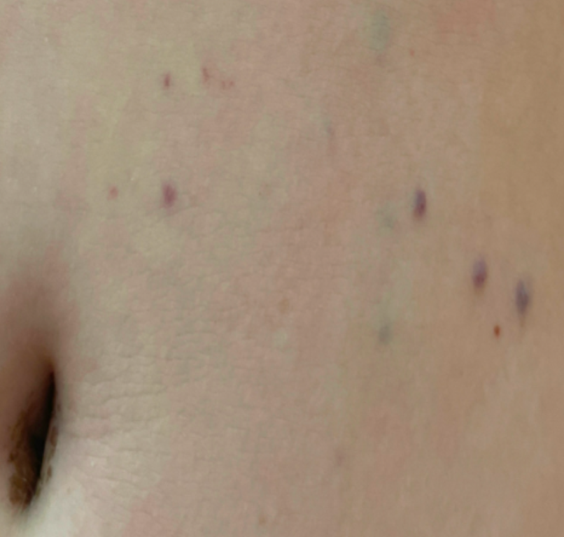
12 yo boy with Crohn's Disease gets IV iron for iron deficiency anemia. His IV infiltrates and discoloration is noticed and persists a year later. What therapy can be offered for this staining? Answer and learn more 👉 bit.ly/3htf9YP?utm_so… #imageofthemonth #quickpoll


12 yo boy developed bluish skin lesions at the neck and abdomen over a few years. Punch biopsy of the skin as shown. No GI complaints. Same lesions in multiple family members. What is the next step? Answer and learn more 👉 bit.ly/3C9ldzo #imageofthemonth #quickpoll


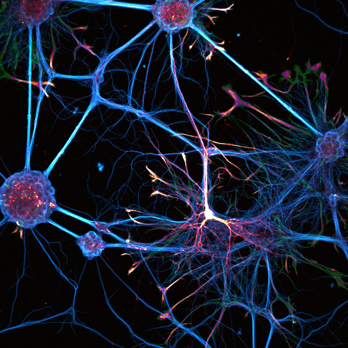
#ImageOftheMonth O.Thoumine @iins_bordeaux & @JTeillon Spheroid made of dissociated rat hippocampal cells observed using confocal after 14 days in culture. A few neurons expressing td-Tomato (coded in gold) have extended long neurites.The spheroid diameter is around 200 µm.

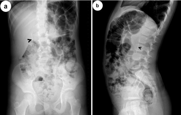
4 yo who swallowed a needle three hours ago goes to the ED. PE is unremarkable. Xray obtained. Where is the ingested needle most likely based on the X-ray? Answer and learn more 👉 bit.ly/3htf9YP?utm_so… #imageofthemonth #quickpoll
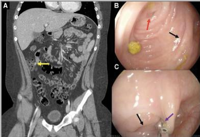
18 mo boy with an esophageal button battery impaction (in situ 26hr). Ultimately, MRA showed moderate mediastinitis and inflammation adjacent to the aorta. How do you follow up with imaging? Answer and learn more 👉 bit.ly/3C9ldzo @alex_s_hudson #imageofthemonth

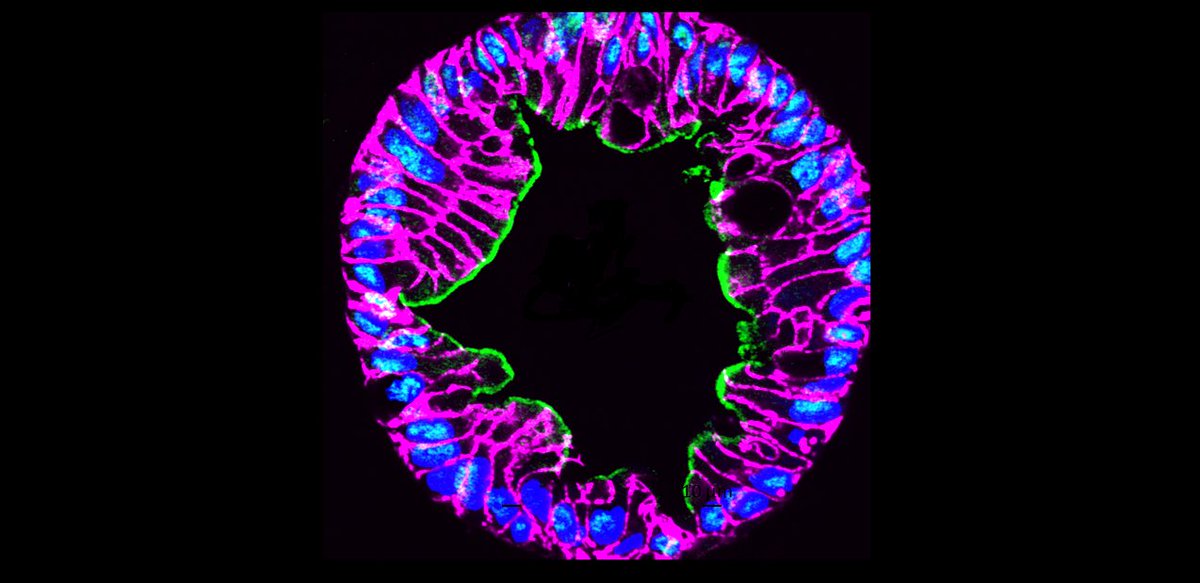
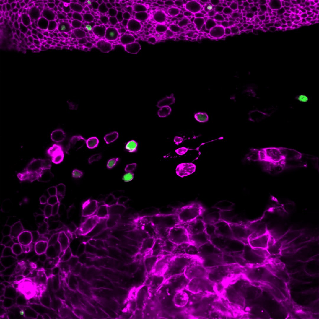
Did you see our January 2024 #ImageoftheMonth? @bcmhouston @EstesLab_BCM, S. Crawford, Z Liu, H. Smith @huntrsmith et al @bcm_gihep #Organoids blogs.bcm.edu/2024/01/02/fro…
12y boy admitted for recurrent abdominal pain leading to restricted food intake. Amylase was 2,254 U/L. No hyperbilirubinemia. Normal US. MRCP was performed. What is the most likely diagnosis? Answer and learn more 👉 bit.ly/3C9ldzo #imageofthemonth #quickpoll

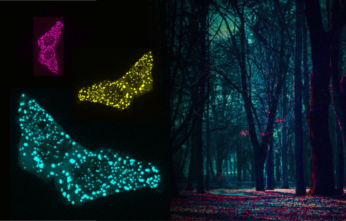
🍁📸 Welcome to our new #ImageOfTheMonth! 🔬✨"Protein Dimerization Butterflies", by @adomostegui, from our Targeted Protein Degradation & Drug Discovery Lab. 👉Protein dimerization obtained by live cell microscopy. Happy #November!🙌 #WeAreIRB
👀Check out our outstanding #ImageOfTheMonth!👇 👁️"The Eye of Sauron", by @KaustuvGhosh1 & David Malpartida, from @MarcoMilanIRB lab. 📸The image shows the pouch of the wing epithelial disc, which gives rise to the wings of adult #Drosophila flies. Happy #September!🌞🍂
✨📸 Introducing our March #ImageoftheMonth! 📸😍 💥"Don't get on my skin nerves!", by @JBonjoch7, @palomasocas and @guiosol, from @AznarLab. 🔍 The image shows the Dermis layer with Schwann cells wrapping the nerves (in green). Happy #March! 🪻🎋🌺 #WeAreIRB

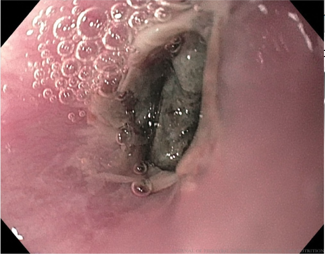
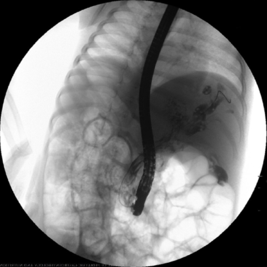
3 yo boy with abd pain and fever for the last 10 days. Abd US shows a dilated CBD and echogenic content. Side viewing endoscopy and cholangiography as shown. What is the next step in management? Answer and learn more 👉 bit.ly/3htf9YP?utm_so… #imageofthemonth #quickpoll
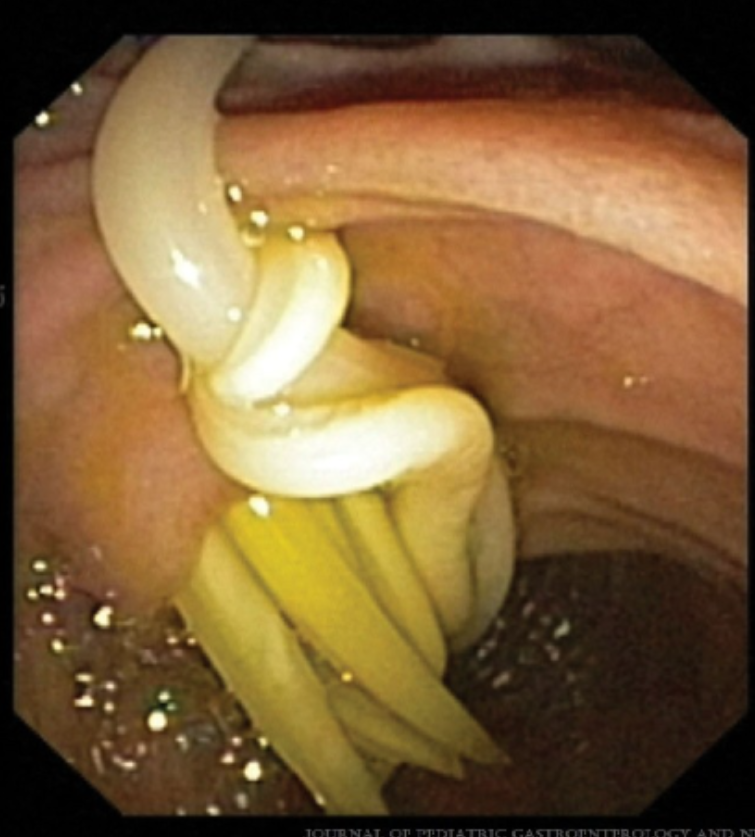
12 yo boy with progressively worsening RLQ pain. CT showing an 11mm structure near the cecum. Colonoscopy shows a cecal diverticula. What is the next best step? Answer and learn more 👉 bit.ly/3htf9YP?utm_so… #imageofthemonth #quickpoll

#ImageoftheMonth 📷 "Chameleons are unique - able to perform large-amplitude, independent eye movements for scanning their environment, then switching to synchronous saccades when tracking prey." Credit: Nima Ghadiri of @LivHospitals

#ImageOfTheMonth This image was acquired by J.Angibaud @iins_bordeaux. Neurons and astrocytes are cultured on a soft polyacrylamide hydrogel. Neurons are labeled for MAP-2 protein (blue), astrocytes for GFAP protein (red) and transfected neurons show GFP expression (green).

🌟New series: "Image of the Month"! Label-free image of a mouse preadipocyte cell!🐭In this cell, organelles were captured at high resolution with Nanolive's 3D Cell Explorer-fluo, using our holotomographic tech. bit.ly/3NVtxJR #Imageofthemonth #Cellimaging #Nanolive

15 yo with abdominal pain and weight loss. CT shows an intraluminal mass in the duodenum. Biopsies shows a predominant spindle cell neoplasm. What is the most common location of this type of tumor? Answer and learn more 👉 #imageofthemonth #quickpoll


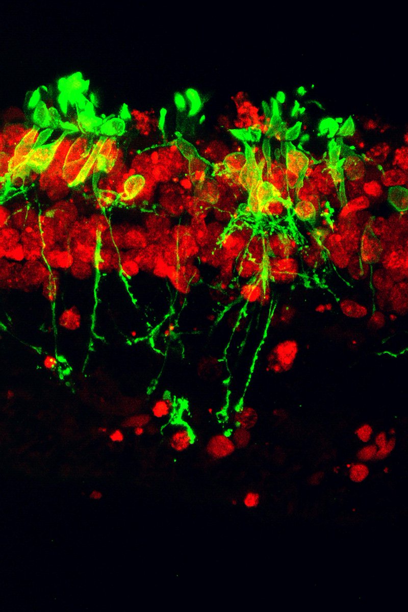
June’s #ImageOfTheMonth is a confocal microscope image of a human retina from an individual with #RetinitisPigmentosa. brnw.ch/21wTFWw #HealthyVision #Visionhealth #Eyes #Science #Research
📸 Introducing our #July #ImageoftheMonth: "Endless Roads of Differentiation"! 🔬By @JBonjoch7, @palomasocas & @guiosol (@aznarlab), this outstanding image shows #immunofluorescence of adult mouse skin, revealing the process of epidermal cell differentiation. 🌞Happy July! 🌊⛱

Do the purple nerve cells in our #ImageOfTheMonth look like sea creatures? 👁🦑 The scientists who took this pic think so! They’re actually called cones—a part of the eye that helps detect color. Check out this image and others in NEI’s image library! brnw.ch/21wW1kg


April is here, and that means a new image of the month, as chosen by the Club President, this months image is “Glacier Lagoon” by Steve Laws. #Photoclub #ImageoftheMonth #StIves #Cambridgeshire

October is here, and that means a new Image of the Month as chosen by the club president - October's image is "Salt pan, Trapani, Sicily" by Stefania Van Lieshout . #Photoclub #ImageoftheMonth #StIves #Cambridgeshire

Do the purple nerve cells in our #ImageOfTheMonth look like sea creatures? 👁🦑 The scientists who took this pic think so! They’re actually called cones—a part of the eye that helps detect color. Check out this image and others in NEI’s image library! brnw.ch/21wW1kg
September’s #ImageOfTheMonth traces the ‘back to school’ phenomenon to the fifth-century BCE. This Greek red-figure cup attributed to the painter Douris prominently displays students, teachers, and the instruments of education. Find out more at peoplingthepast.com!

September is here, and a new Image of the Month as chosen by the club president - September's image is “Norfolk Reflections" by Nigel Sandbrook. #Photoclub #ImageoftheMonth #StIves #Cambridgeshire
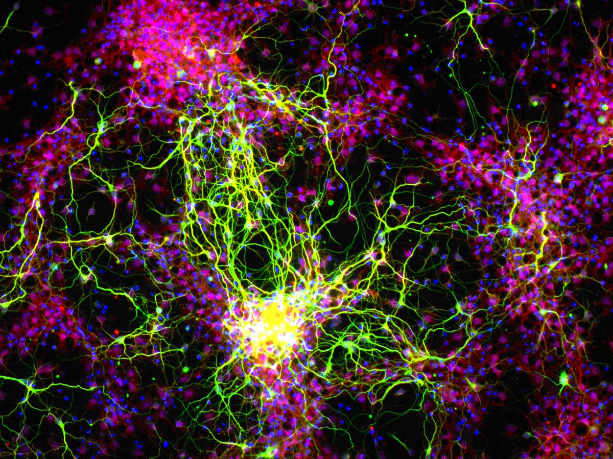
Drumroll, please!! Introducing our #ImageOfTheMonth for August. This photo shows new neurons (green) and their supporting cells, called astrocytes (red), created in a petri dish from stem cells. brnw.ch/21wUTEs #Vision
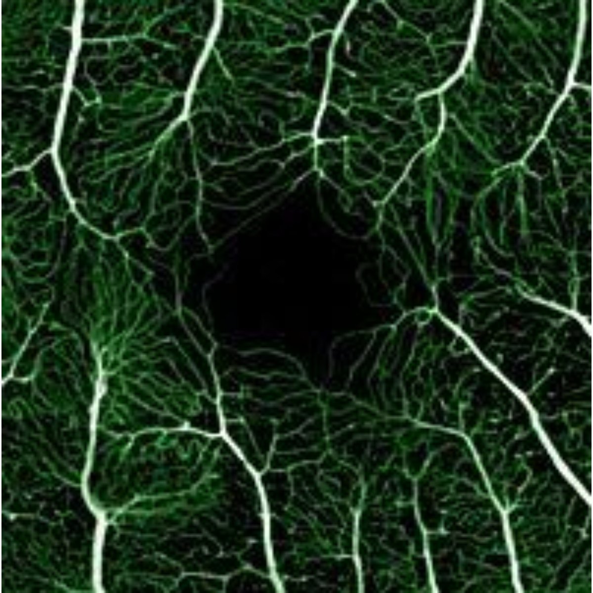
Check out our #ImageoftheMonth for a peek at the blood vessels surrounding the center of the retina (the part of your eye responsible for sharp, central vision. These tiny branches play a big role in helping you read, drive, and recognize faces! brnw.ch/21wUEnp

August has arrived, and a new Image of the Month as chosen by the club president - August's image is “Inveraray Harbour# by Stu Hames. #Photoclub #ImageoftheMonth #StIves #Cambridgeshire

July’s image of the month features branching blood vessels that surround the central part of a retina. For ease of viewing, the blood vessels have been lit up green by fluorescein angiography combined with adaptive optics. brnw.ch/21wUbT7 #ImageOfTheMonth
July is here already! A new Image of the Month as chosen by the club president - July's image is “Remembrance ” by David King. #Photoclub #ImageoftheMonth #StIves #Cambridgeshire

June’s #ImageOfTheMonth is a confocal microscope image of a human retina from an individual with #RetinitisPigmentosa. brnw.ch/21wTFWw #HealthyVision #Visionhealth #Eyes #Science #Research
June’s #ImageOfTheMonth is a confocal microscope image of a human retina from an individual with #RetinitisPigmentosa. brnw.ch/21wTFWt #HealthyVision #Visionhealth #Eyes #Science #Research


June's Microscopy #ImageOfTheMonth features a heart-shaped multicellular spheroid formed by lung tumour cells, fibroblasts and macrophages💚🔬 📸 Marina Tuxans, from the Lung Immunity Translational research group flic.kr/p/2rasYUW #IGTPMicroscopy


#imageofthemonth Out of the Woods: Reversing a Trend of Increased ICU Readmissions in Trauma Patients. This cover image art was created by Shelby Osbourn, Graphic Artist at the University of Arkansas for Medical Sciences Institute for Community Health Innovation.

June is here, and so it's time for a new Image of the Month as chosen by the club president - June's image is “The shiniest jag in the carpark” by Angels Lucas. #Photoclub #ImageoftheMonth #StIves #Cambridgeshire


Showcasing the beauty in the little things 🎨🔬 Our #ImageOfTheMonth comes from Rakshitha Ravikumar: “Each cell is drawn with care, curiosity, and a splash of colour… plus a panda who just had to be part of it.” 👉 ow.ly/Mp5F50VWryN #PathArt #Histology #ThePathologist

In honor of Mother’s Day on May 11th, our #ImageOfTheMonth is a hydria (water jug) depicting a mother handing her infant to a nursemaid. The domesticity of this scene is typical of High Classical Greece. Dating to c.440–430 BCE, this vessel is now in the Harvard Art Museum.

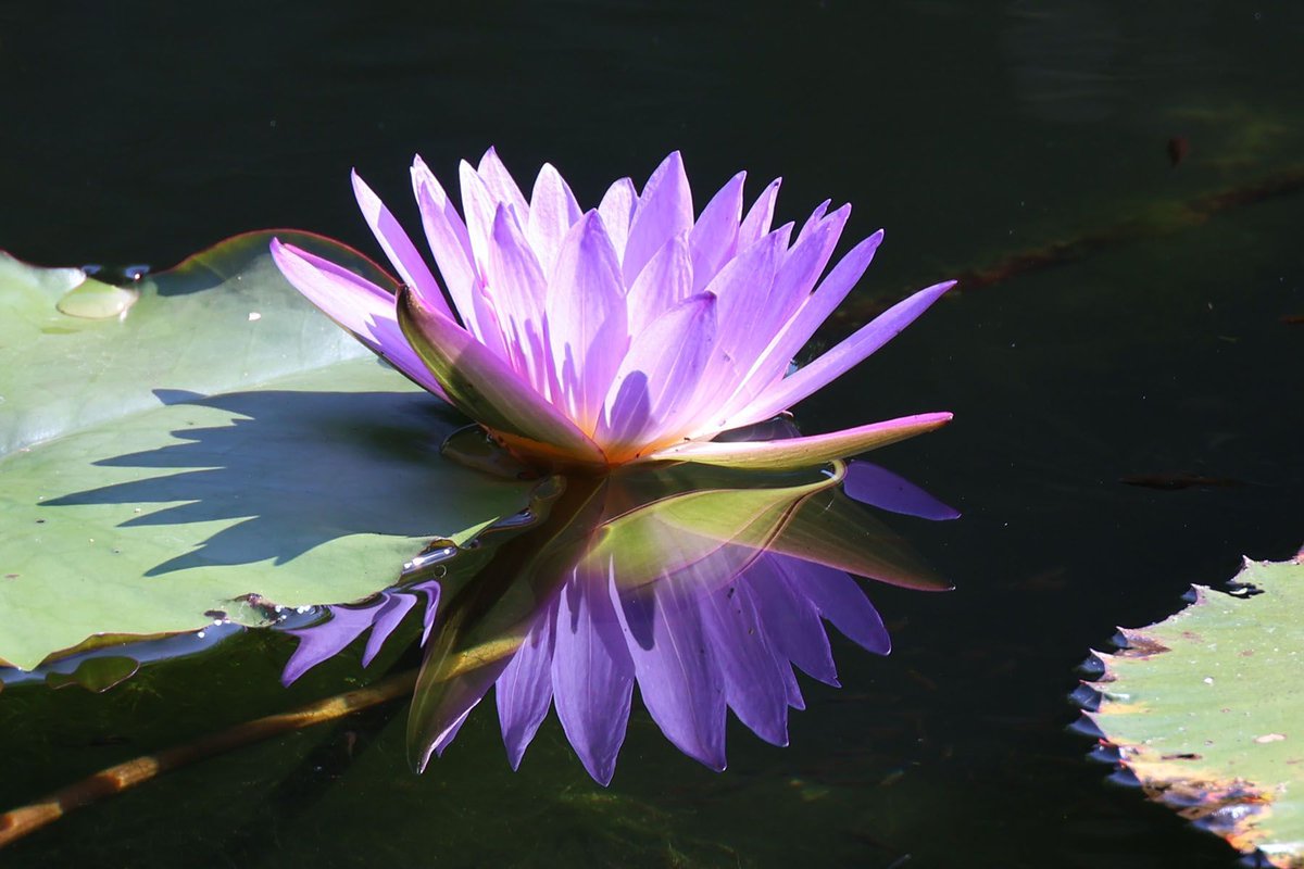
May has arrived and so has a new Image of the Month as chosen by the club president - May's image is “Water Lily” by Sarah Bassett. #Photoclub #ImageoftheMonth #StIves #Cambridgeshire

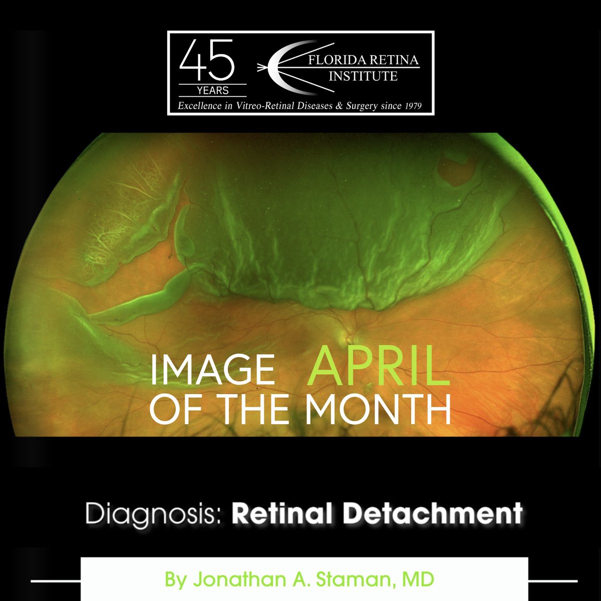
Florida Retina Institute’s April Image of the Month. Diagnosis: Retinal Detachment, Captured by Dr. Jonathan A. Staman, this powerful image highlights the urgency of expert retina care. Learn more at floridaretinainstitute.com #retinaldetachment #imageofthemonth #eyehealth
Make way! April’s #ImageOfTheMonth provides a close-up look at a microglial cell (green) as it extends spider-like arms to capture and consume rod photoreceptor cells (blue). medialibrary.nei.nih.gov/search?keyword…

In honor of #InternationalWomensDay, our #ImageOfTheMonth is a marble relief commemorating two female gladiators —Amazon and Achillia. Found in Halicarnassus, this relief was acquired through donation by the @britishmuseum (1847,0424.19) in 1846, and prior provenience is murky.

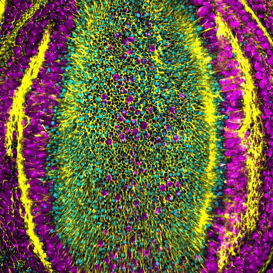
🌺🌞June has arrived, and so has our #ImageoftheMonth: "The Silk Road of Hematopoiesis" by Daniel Fernández from @FraticelliLab. 📸 Featuring scRNA-seq of single bone marrow cells from adult mice transplanted with embryonic or adult hematopoietic stem cells. Happy #June! ✨
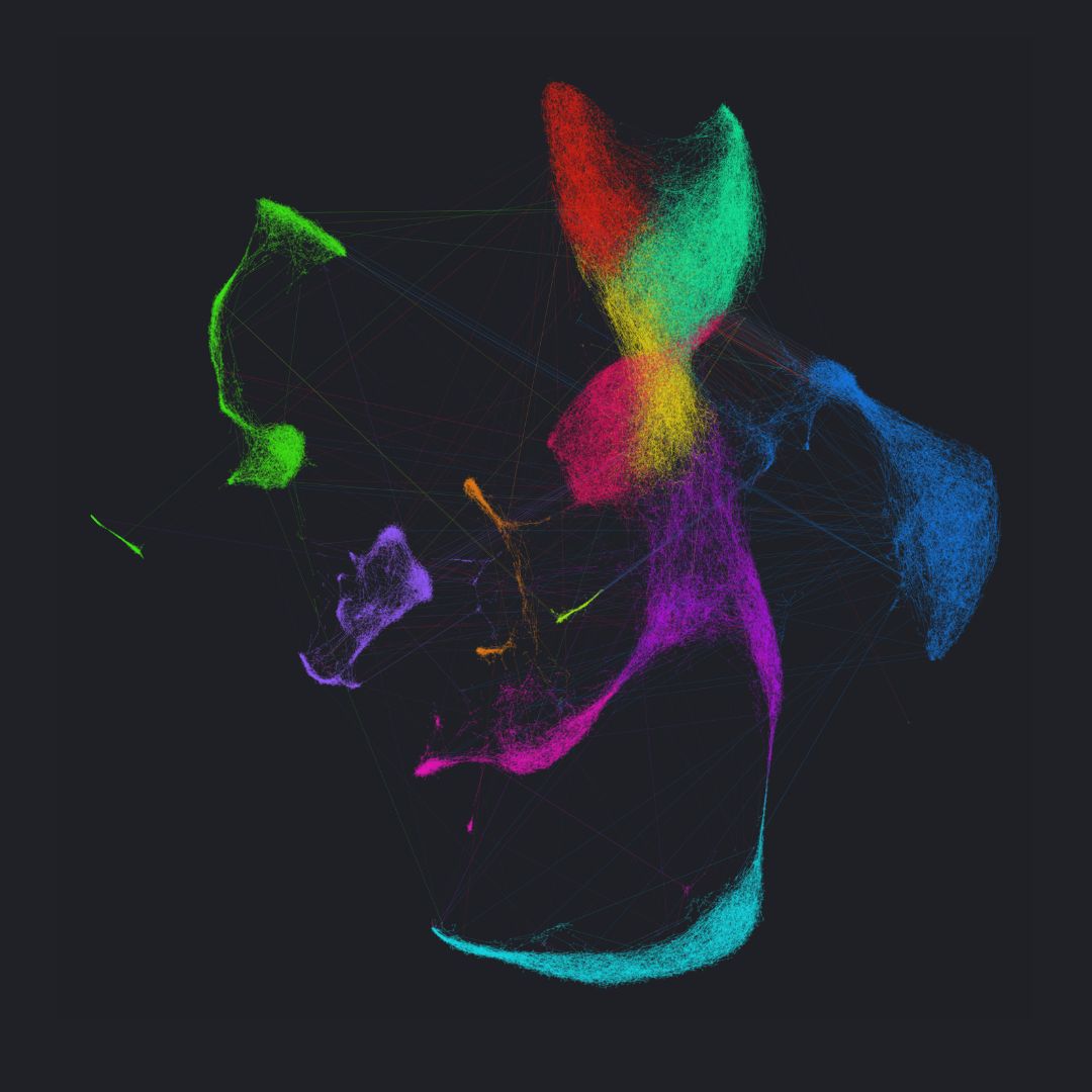
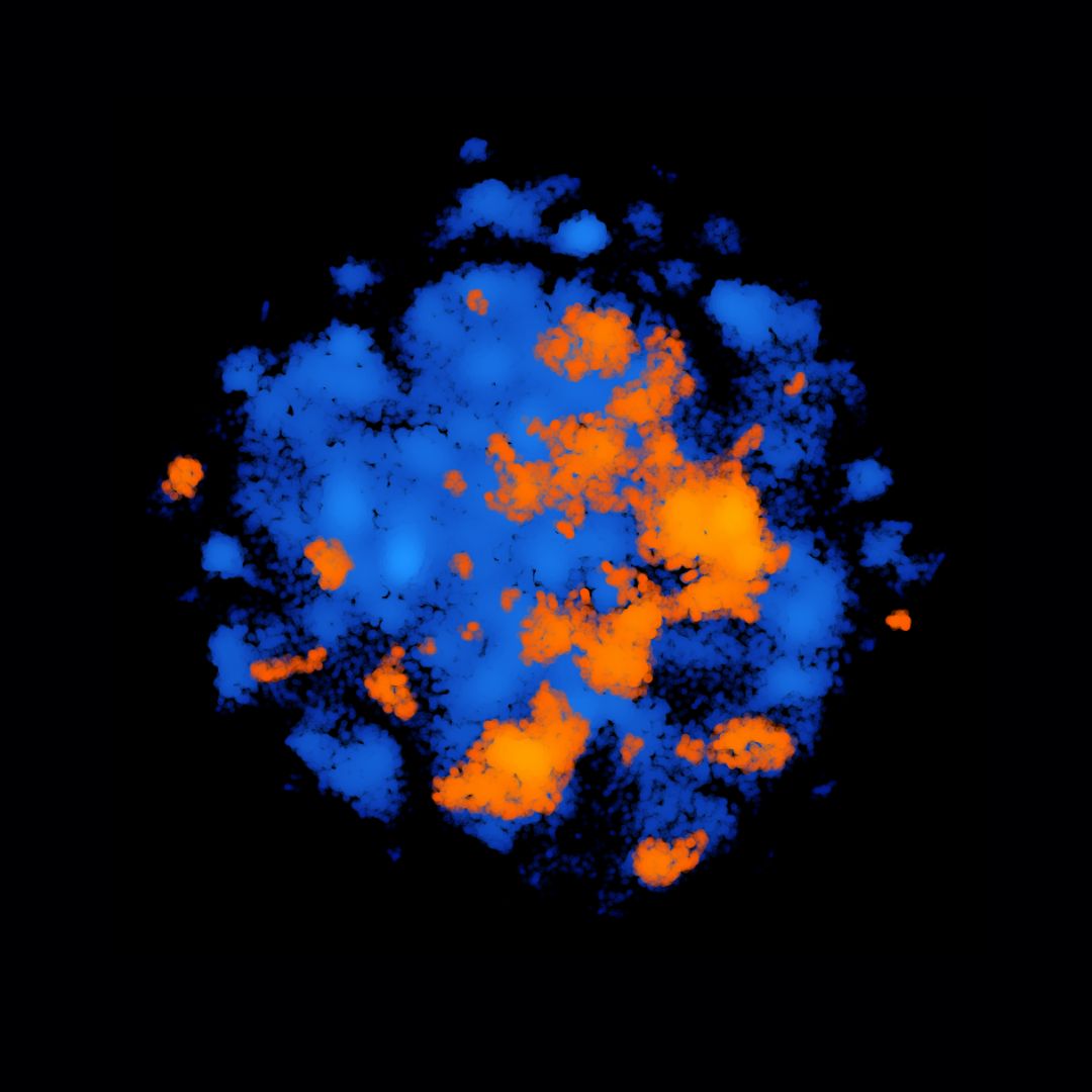
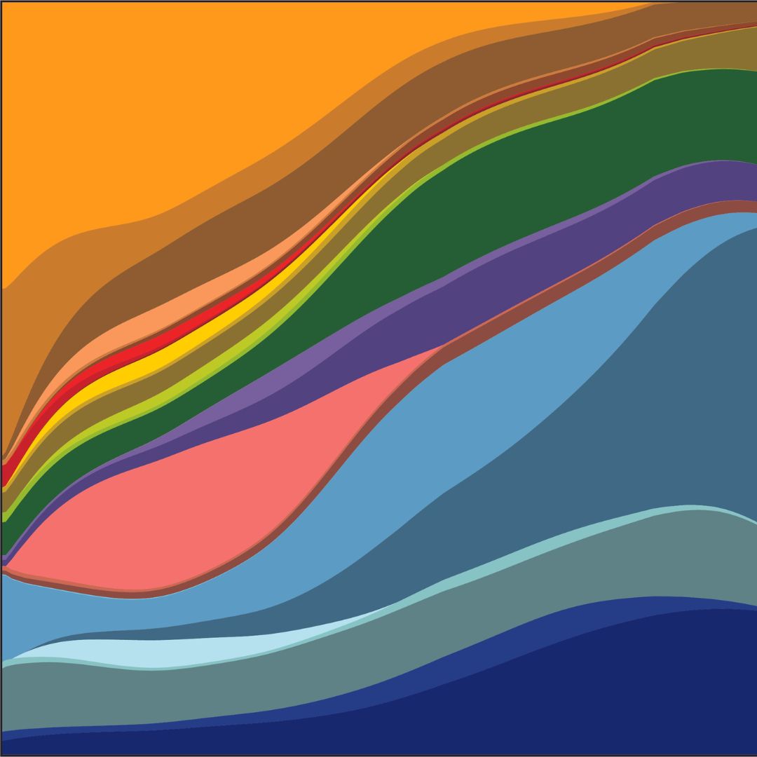
🍂Welcoming #October with this amazing #ImageOfTheMonth! 🧫"Molecular Clouds" is a representation of the old #IRBBarcelona Chemical Library (🟠orange), together with the new one (🔵blue). 📸@acomajuncosa et al. (Structural Bioinformatics & Network Biology, led by Dr. @ptck72).
January 2026 is here, and a new month means a new image of the month, as chosen by the Club President, this months image is “Grey Seal Pup looking for Mum ” by Gary Dean. #Photoclub #ImageoftheMonth #StIves #Cambridgeshire

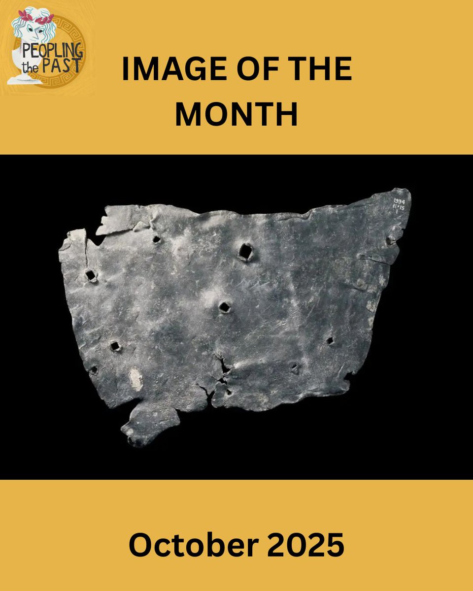
In the spirit of Halloween, October’s #ImageOfTheMonth is a particularly cursed item: a Roman curse tablet (defixio) from 1st–4th century CE London (British Museum 1934,1105.1). This thin sheet of lead wishes ill-health to a woman named Tretia Maria /1
👀Check out our outstanding #ImageOfTheMonth!👇 🌴🌬️ It's "Breathing Trunks", by Panagiotis Giannios, from @MarcoMilanIRB lab. 🖼️🪰A captivating composition of the respiratory structures of #Drosophila melanogaster, with cell nuclei in ⚫️black. Happy #August everyone!🌞

🌞Let's start #February with this fascinating #ImageoftheMonth, entitled "Mitochondrial Cat's Eyes"! 👀🐈 📸By Nickolaos Giakoumakis, Chong Zhang & Paula Sánchez, it image shows the contours of segmented #mitochondria with impressive precision. Happy February everyone! ☔️🤍

#ImageOfTheMonth July image from @ang_getz @iins_bordeaux shows a sparse labeling of endogenous AMPA receptors on CA1 pyramidal neuron dendrites in the stratum radiatum. 3D image was acquired using lattice light-sheet microscopy.

#ImageOfTheMonth This image was acquired by J.Angibaud @iins_bordeaux. Neurons and astrocytes are cultured on a soft polyacrylamide hydrogel. Neurons are labeled for MAP-2 protein (blue), astrocytes for GFAP protein (red) and transfected neurons show GFP expression (green).
#ImageoftheMonth 📷 "Chameleons are unique - able to perform large-amplitude, independent eye movements for scanning their environment, then switching to synchronous saccades when tracking prey." Credit: Nima Ghadiri of @LivHospitals

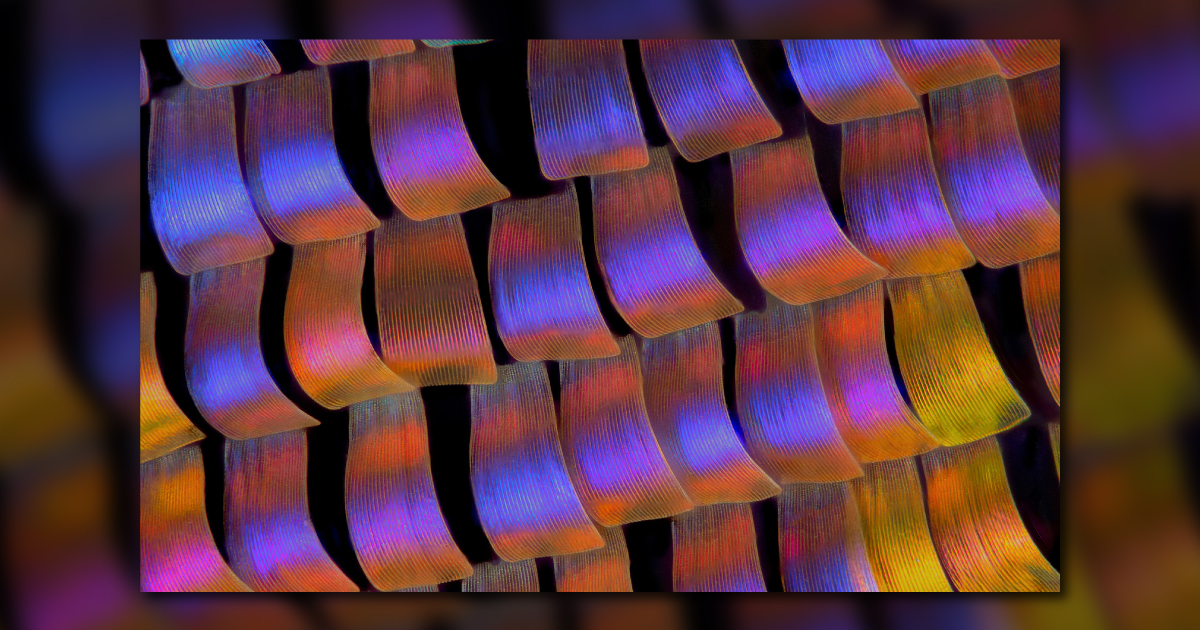
🤩 Behold September's #ImageoftheMonth: Twilight Wings! 🦋 This stunning shot of the twilight moth Urania ripheus reveals its enchanting wing scales. 📸✨ Honorable Mention winner in Olympus/Evident Image of the Year Award 2020 contest. Courtesy of Walter Ferrari of Argentina.
📸 October's #ImageoftheMonth! This stunning image features a close-up view of 431 stainless steel, etched with modified Murakami's technique at a magnification of 400X using digital image correlation. Courtesy of Pace Technologies. #MaterialsMonday #MaterialsScience #Microscopy
September’s #ImageOfTheMonth traces the ‘back to school’ phenomenon to the fifth-century BCE. This Greek red-figure cup attributed to the painter Douris prominently displays students, teachers, and the instruments of education. Find out more at peoplingthepast.com!
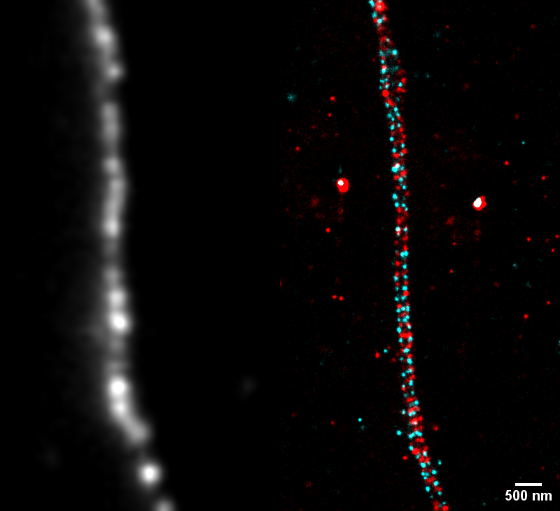
#ImageOftheMonth Xuesi Zhou @iins_bordeaux Adducin (cyan) and β2-spectrin (red), are imaged using exchange-PAINT super-resolution microscopy. One can see the alternative 90nm ring-like structures in neurites. Left image shows the neurite using a MAP2 staining.
✨📸 Introducing our March #ImageoftheMonth! 📸😍 💥"Don't get on my skin nerves!", by @JBonjoch7, @palomasocas and @guiosol, from @AznarLab. 🔍 The image shows the Dermis layer with Schwann cells wrapping the nerves (in green). Happy #March! 🪻🎋🌺 #WeAreIRB
#ImageOftheMonth Hélène Bonnet @INRAE_BFP In June say it with flowers. This image, taken under a macroscope at the plant unit of the BIC, shows flower bud from the sweet cherry tree ‘Burlat’, sampled in the orchard and grown at 24°C for ten days to observe flowering.

Make way! April’s #ImageOfTheMonth provides a close-up look at a microglial cell (green) as it extends spider-like arms to capture and consume rod photoreceptor cells (blue). medialibrary.nei.nih.gov/search?keyword…
❄️Le'ts celebrate #February with this 🔝 #ImageoftheMonth from our calendar! 📸"Breaching Boundaries", by @KaustuvGhosh1, from @MarcoMilanIRB's lab. 🔬Wild-type epithelial cells (upper) & tumorigenic cells (lower), membranes in🟣magenta & caspase reporter in🟢green. #WeAreIRB
It’s our October #ImageOfTheMonth! This photo features retinal pigment epithelial cells stained red by RPE65 antibody. RPE65 gene mutations can cause a form of Leber congenital amaurosis (LCA), a disease that leads to visual impairment early in childhood. bit.ly/3YeJnFi
🐪🌅 Like walking through cellular dunes, here comes our #ImageoftheMonth, by @adriacanyellas, from @BatlleLab. 📸 "Cellular Dunes" shows the evolution of the tumor microenvironment during metastatic growth in the liver (from left to right). Happy #August!🌞🏖
Something went wrong.
Something went wrong.
United States Trends
- 1. #WrestleMania N/A
- 2. Luke Kennard N/A
- 3. Bengals N/A
- 4. Lakers N/A
- 5. #LakeShow N/A
- 6. Rockets N/A
- 7. #ImmortalCF N/A
- 8. #Toonami N/A
- 9. Sengun N/A
- 10. Giants N/A
- 11. Dexter Lawrence N/A
- 12. Porter Martone N/A
- 13. #TokyoRevengers N/A
- 14. Orton N/A
- 15. Bianca N/A
- 16. Flyers N/A
- 17. Paige N/A
- 18. Bron N/A
- 19. Pat McAfee N/A
- 20. Trey N/A