#scanningelectronmicroscope search results

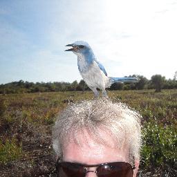
The new type SEM has arrived at the Wakamiya Lab. It will show us many new worlds✨😊✨ #SU8600 #Hitachi #ScanningElectronMicroscope


Image of the Day ~ Clogging Bricks: SEM image of Calcium carbonate crystals. CREDIT: Fabio Dossi, ITP; JSM-IT200 #ScanningElectronMicroscope #ImageContest

Nice try but #nocigar -- when you need a #scanningelectronmicroscope to see who the #USMint is honoring on #thisquarter

Working with a Scanning Electron Microscope | 2009 I will mint some images made with electrons in the near future. #SEM #ScanningElectronMicroscope #ArtScience #MicroMeter #Scale #HiddenWorlds

Image of the Day #3 ~ Countdown for the 2023 JEOL #ImageContest: "Green Tadpoles" - Chlamydospores (resistance structures) of Ganoderma sp.; CREDIT: Jander Matos Guimarães, Aldenora dos Santos, CMABio/UEA; JSM IT500HR #ScanningElectronMicroscope

Who knew there was a #worm23 conference? We're jumping in to share a couple of STEM images of C. elegans taken on our #NeoScope benchtop SEM! #scanningelectronmicroscope #nematode #microscopy


Image of the Day ~ "Clusters of Grapes" Soot Particles and Carbonate Crystals. CREDIT: Brandon Aldinger, American Glass Research; JEOL IT-300LV #ScanningElectronMicroscope


A cool guide to what goes on inside a scanning electron microscope. #coolguides #ScanningElectronMicroscope #SEM #Microscopy #Science

It's #MicroscopyMonday! "High stellar resolution" shows residual sodium crystal in endodontic rasp; CREDIT: Jander Matos Guimarães and Jéssica Araújo Marques Matos Guimarães,CMABio/UEA; JSM IT500HR #ScanningElectronMicroscope

We're demonstrating the new IT710HR LV Field Emission SEM at #MM2023. The hybrid Secondary Electron Detector with photon detection can be used as a panchromatic CL detector, in this image showing trace element zoning in Zircon. #microscopy #scanningelectronmicroscope


"Invaded Space" by Jander Matos Guimarães, UNIVERSIDADE DO ESTADO DO AMAZONAS wins the JEOL August Image Contest. Bacterial biofilm from the egg surface of Rhodnius prolixus (infectious agent from chagas disease). JSM IT500HR #ScanningElectronMicroscope.

Our Y2 students visited our newly refurbished microscopy lab to see our scanning electron microscope. Our lab manager Sharon Gibbons was showing our students about the wide range of samples that can be fitted into the microscope @RoyalHolloway #scanningelectronmicroscope


📹 Don’t worry if you missed our recent Tutorial, “Introduction to sample identification in the SEM with AZtecMatch” you can now watch it on demand here 👉 okt.to/5HqREX #SampleIdentification #ScanningElectronMicroscope


Embarking on a microscopic journey, our SEM-EDS technology at @creativenano is unraveling the secrets of #microplastics . Take a moment to observe the compelling #scanningelectronmicroscope (#SEM) image of a microplastic captured by our team!


Introducing #JEOL's JSM-IT210 and JSM-IT710HR Scanning Electron Microscopes! 🔬 Click here for more info ℹ lnkd.in/gVHF9-d7 #scanningelectronmicroscope #microscopy #SEMA
Image of the Day ~ Alumina silicate after reaction with 0.1M sodium hydroxide at reflux for 5 hours; CREDIT: Marcio de Paula, University of São Paulo; JEOL JSM-7200F SEM. #ScanningElectronMicroscope #MaterialsScience #ImageContest


Microlite hunting today in some Tephra from the 1902 St Vincent eruption. It’s my final session getting to use the SEM in the Plymouth Electron Microscopy Centre and I’m definitely going to miss it. #SEM #ScanningElectronMicroscope #Microlites #Volcanology #MastersThesis


Read the full article 👉ieeexplore.ieee.org/abstract/docum… Well done Abdallah Alakayleh and team 👏 #scanningelectronmicroscope #ImageAnalysis #electricaldevices
Image of the Day ~ Sand Dollar by Sheri Neva, Eurofins EAG Los Angeles, taken on JEOL JSM-6610LV SEM. Beautiful detail of this exoskeleton #microscopy #scanningelectronmicroscope #sanddollar

Image of the Day ~ in honor of the Florida Atlantic Men's #Basketball spectacular season @FAUMBB the @fauhs_research Owls Lab imaged the leather of a basketball in their NeoScope Benchtop SEM. #Microscopy #ScanningElectronMicroscope

Read the full article 👉ieeexplore.ieee.org/abstract/docum… Well done Abdallah Alakayleh and team 👏 #scanningelectronmicroscope #ImageAnalysis #electricaldevices
Working with a Scanning Electron Microscope | 2009 I will mint some images made with electrons in the near future. #SEM #ScanningElectronMicroscope #ArtScience #MicroMeter #Scale #HiddenWorlds

कार्यक्रम प्रतिभागियों को एमआरसी के तकनीकी कर्मचारियों की देखरेख में माइक्रोस्कोप के संचालन का व्यावहारिक अनुभव प्रदान करेगा। #ScanningElectronMicroscope #SEM #TrainingProgram #MaterialScience #Research #LabTraining #MNITJaipur #TechnicalSkills #HandsOnExperience
SEM Machines Installed on Dynemech Anti-Vibration Tables To learn more: vibrationmountsindia.com/products/ 📞+91-9810760131 | +91-9911145131 📧[email protected] 🌐vibrationmountsindia.com|https://t.co/5ieoe9SLF5 #ScanningElectronMicroscope #AntiVibrationTable #DynemechSolutions#SEMStability

Our Y2 students visited our newly refurbished microscopy lab to see our scanning electron microscope. Our lab manager Sharon Gibbons was showing our students about the wide range of samples that can be fitted into the microscope @RoyalHolloway #scanningelectronmicroscope
Nice try but #nocigar -- when you need a #scanningelectronmicroscope to see who the #USMint is honoring on #thisquarter

Research Staff (f_m_x) in Scanning Electron Microscopy (SEM) - Potsdam, Germany - earthworks-jobs.com/jobs/gfz24059 - #jobs #research #scanningelectronmicroscope #SEM #mineralogy #biogeochemistry #materialsscience #geoscience
Image of the Day ~ "Clusters of Grapes" Soot Particles and Carbonate Crystals. CREDIT: Brandon Aldinger, American Glass Research; JEOL IT-300LV #ScanningElectronMicroscope
Happy Spring! Today our #ImageoftheDay is "The Hatching". In this SEM image it's not an egg, but a #silver bubble imaged by Ajitzi Negrete at Stratascan using a JSM-IT510 #scanningelectronmicroscope. Join in the fun and enter the JEOL 2024 #ImageContest bit.ly/3vNzHG3

Embarking on a microscopic journey, our SEM-EDS technology at @creativenano is unraveling the secrets of #microplastics . Take a moment to observe the compelling #scanningelectronmicroscope (#SEM) image of a microplastic captured by our team!
Image of the Day #3 ~ Countdown for the 2023 JEOL #ImageContest: "Green Tadpoles" - Chlamydospores (resistance structures) of Ganoderma sp.; CREDIT: Jander Matos Guimarães, Aldenora dos Santos, CMABio/UEA; JSM IT500HR #ScanningElectronMicroscope

Dive into the World of Discovery with the Latest in #Microscope Technology Read: alliedmarketresearch.com/microscope-mar… Uncover the secrets of the unseen and explore the Microscope Market trends that are reshaping science. #MicroscopeMarket #ScanningElectronMicroscope #ElectronMicroscope
alliedmarketresearch.com
Microscope Market Size, Share, Growth & Report - 2027
The microscope market was valued at $1.39 billion in 2019 and is expected to reach $1.64 billion by 2027; The market is divided by type, end use, and region.
‘Through the Lens of Progress: Unveiling the Dynamics of the Microscope Market’ Read: link.medium.com/2HDmE4Q8TEb #Microscope #MicroscopeMarket #ScanningElectronMicroscope #Microscoy #ElectronMicroscope
link.medium.com
“Through the Lens of Progress: Unveiling the Dynamics of the Microscope Market”
In the realm of scientific exploration, the microscope stands as an unassuming yet indispensable tool that has fueled countless…

The @thermofisher Perception Software offers automated analysis, classification, and reporting that turns the #ScanningElectronMicroscope of choice into a dedicated #ParticleAnalysis solution. Read more in the latest blog: ter.li/30lngk

The new type SEM has arrived at the Wakamiya Lab. It will show us many new worlds✨😊✨ #SU8600 #Hitachi #ScanningElectronMicroscope
Who knew there was a #worm23 conference? We're jumping in to share a couple of STEM images of C. elegans taken on our #NeoScope benchtop SEM! #scanningelectronmicroscope #nematode #microscopy
Nice try but #nocigar -- when you need a #scanningelectronmicroscope to see who the #USMint is honoring on #thisquarter
Image of the Day ~ Clogging Bricks: SEM image of Calcium carbonate crystals. CREDIT: Fabio Dossi, ITP; JSM-IT200 #ScanningElectronMicroscope #ImageContest
It's #MicroscopyMonday! "High stellar resolution" shows residual sodium crystal in endodontic rasp; CREDIT: Jander Matos Guimarães and Jéssica Araújo Marques Matos Guimarães,CMABio/UEA; JSM IT500HR #ScanningElectronMicroscope
We're demonstrating the new IT710HR LV Field Emission SEM at #MM2023. The hybrid Secondary Electron Detector with photon detection can be used as a panchromatic CL detector, in this image showing trace element zoning in Zircon. #microscopy #scanningelectronmicroscope
Image of the Day ~ "Clusters of Grapes" Soot Particles and Carbonate Crystals. CREDIT: Brandon Aldinger, American Glass Research; JEOL IT-300LV #ScanningElectronMicroscope
Happy Spring! Since today is #NationalDaffodilDay we took a closer look at daffodil #pollen with our #ScanningElectronMicroscope. bit.ly/3qTofSS


Image of the Day #3 ~ Countdown for the 2023 JEOL #ImageContest: "Green Tadpoles" - Chlamydospores (resistance structures) of Ganoderma sp.; CREDIT: Jander Matos Guimarães, Aldenora dos Santos, CMABio/UEA; JSM IT500HR #ScanningElectronMicroscope
Image of the Day ~ Alumina silicate after reaction with 0.1M sodium hydroxide at reflux for 5 hours; CREDIT: Marcio de Paula, University of São Paulo; JEOL JSM-7200F SEM. #ScanningElectronMicroscope #MaterialsScience #ImageContest
Image of the Day ~ in honor of the Florida Atlantic Men's #Basketball spectacular season @FAUMBB the @fauhs_research Owls Lab imaged the leather of a basketball in their NeoScope Benchtop SEM. #Microscopy #ScanningElectronMicroscope
A cool guide to what goes on inside a scanning electron microscope. #coolguides #ScanningElectronMicroscope #SEM #Microscopy #Science
Image of the Day ~ Sand Dollar by Sheri Neva, Eurofins EAG Los Angeles, taken on JEOL JSM-6610LV SEM. Beautiful detail of this exoskeleton #microscopy #scanningelectronmicroscope #sanddollar
Image of the Day ~ "Life Beyond the Wall" - L-forms escaping antibiotic repression. CREDIT: Joost Willemse, Leiden University. JEOL JSM-7600F #ScanningElectronMicroscope #cellbiology #bacteria #microscopy

Image of the Day ~ "Marine Flowers" Calcareous #nanoplankton from the Southern Indian Ocean.; CREDIT: Sahina Gazi, National Centre for Polar and Ocean Research (NCPOR), Goa; JEOL JSM-7610F FE-SEM #scanningelectronmicroscope #oceanography. #ImageContest bit.ly/3Hil2EW

"Invaded Space" by Jander Matos Guimarães, UNIVERSIDADE DO ESTADO DO AMAZONAS wins the JEOL August Image Contest. Bacterial biofilm from the egg surface of Rhodnius prolixus (infectious agent from chagas disease). JSM IT500HR #ScanningElectronMicroscope.
Image of the Day ~ "Invaded space" Bacterial biofilm from egg surface of Rhodnius prolixus (infectious agent from chagas disease); CREDIT: Jander Matos Guimarães, UNIVERSIDADE DO ESTADO DO AMAZONAS. Imaged on JEOL JSM-IT500HR #SEM #ScanningElectronMicroscope #bacteria #microscopy

SEM Machines Installed on Dynemech Anti-Vibration Tables To learn more: vibrationmountsindia.com/products/ 📞+91-9810760131 | +91-9911145131 📧[email protected] 🌐vibrationmountsindia.com|https://t.co/5ieoe9SLF5 #ScanningElectronMicroscope #AntiVibrationTable #DynemechSolutions#SEMStability
Working with a Scanning Electron Microscope | 2009 I will mint some images made with electrons in the near future. #SEM #ScanningElectronMicroscope #ArtScience #MicroMeter #Scale #HiddenWorlds
📹 Don’t worry if you missed our recent Tutorial, “Introduction to sample identification in the SEM with AZtecMatch” you can now watch it on demand here 👉 okt.to/5HqREX #SampleIdentification #ScanningElectronMicroscope
Something went wrong.
Something went wrong.
United States Trends
- 1. Broncos 63.1K posts
- 2. Bo Nix 17.5K posts
- 3. yeonjun 165K posts
- 4. Geno 17.8K posts
- 5. $SMILEY N/A
- 6. Sean Payton 4,595 posts
- 7. #TNFonPrime 3,952 posts
- 8. Kenny Pickett 1,488 posts
- 9. DANIELA 32.6K posts
- 10. #NOLABELS_PART01 67.8K posts
- 11. Chip Kelly 1,928 posts
- 12. Jalen Green 6,853 posts
- 13. Bradley Beal 3,259 posts
- 14. NO LABELS NOVEMBER 23K posts
- 15. Pete Carroll 1,841 posts
- 16. #criticalrolespoilers 4,302 posts
- 17. TALK TO YOU OUT NOW 23.1K posts
- 18. Troy Franklin 2,390 posts
- 19. Jeanty 6,434 posts
- 20. byers 27.7K posts