#cellimaging kết quả tìm kiếm
Seeing cytotoxicity in full color 🎨 Nanolive’s LIVE Cytotoxicity Assay reveals how compounds and concentrations shape cancer cell death dynamics in real time. 👉 bit.ly/3KmBOrk #DrugDiscovery #Cytotoxicity #CellImaging

.@WeillCornell researchers have developed a new pH sensor, the Acidic pH Indicator Dye, with improved function in the physiological pH range of late endosomes & lysosomes. Learn more: lnkd.in/efqj4ibi #AlzheimersResearch #Neurodegeneration #cellimaging #Fluorescence


🧫🔬 Preserve cell health and minimize background noise during extended ambient exposure with a clear, physiological buffer. Try Live Cell Imaging Solution for reliable, high-quality imaging results - spr.ly/60177G9t9 #EvosEasy #WeeklyTip #CellImaging

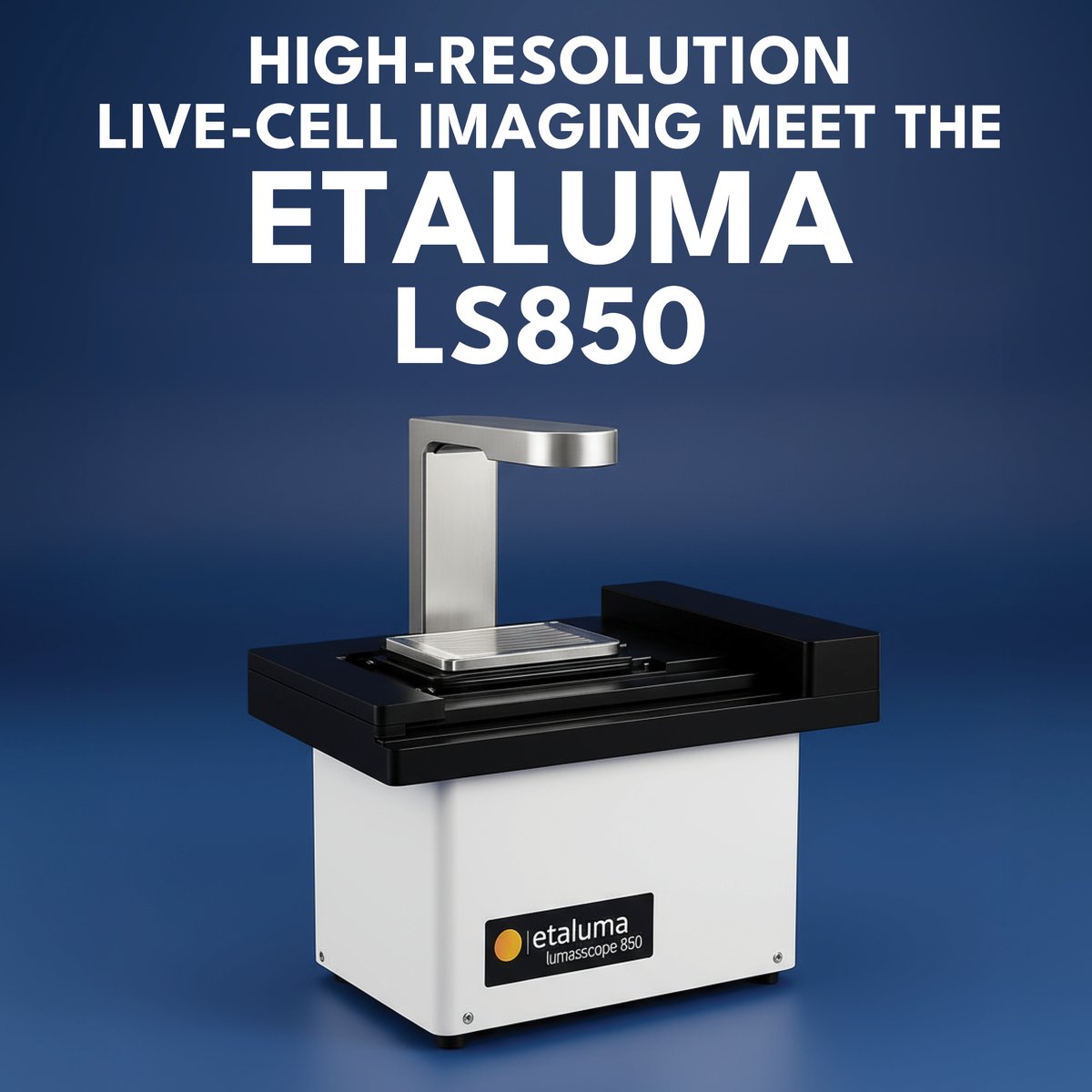
Explore the Lumascope 850™ from @Etaluma — available now at FroggaBio! Compact yet potent, this device merges high-resolution imaging with automated functionality for seamless live cell imaging.🐸🔬💡 LEARN MORE: hubs.li/Q03MwngT0 #Microscopy #CellImaging #Lab #FroggaBio
📊 Avoid 96-well plates with offset feet to minimize environmental disturbances. Choose the right vessel for your sample to ensure reliable imaging results. 🧫 spr.ly/60137G9xz #EvosEasy #WeeklyTip #CellImaging #LabEfficiency


Welcome to the new members of our community! 🌟Nanolive's high-res images and analysis offer unique cellular insights. Our holotomographic microscopes, with a patented rotating light source deliver high-res 3D images. bit.ly/41Xbl8I #Nanolive #CellImaging #Innovation

Greetings, people! Quick Q: Is there any open-source tool to analyze quantitative phase imaging (QPI) data? Please let me know. Thanks. #microscopy #cellimaging #quantitativephaseimaging #cellmass #celldensity 🔬


An overnight timelapse showing the stunning view of budding snowflake yeast, captured through label-free live cell imaging with the @Tomocubeinc HT-X1 #Holotomography system. #livecell #cellimaging #3Dimaging #microscopy #labelfree
📸Unveil the effects of phototoxicity on WM793 melanoma cells with Nanolive's 3D Cell Explorer-fluo! Imaged every 30s for 6h, witness the impact of phototoxicity on cell and organelle structures. bit.ly/3vGkKFx #Phototoxicity #CellImaging #Nanolive
🔬 Witness the magic of stem cell division! Our label-free imaging captures their journey for 15 hours straight, unveiling insights into their vitality and behavior. More info on stem cell research with label-free imaging: bit.ly/49C7a4I #StemCells #CellImaging #Nanolive
Dive into the secret life of your skin!✨ 90% of skin's outer cells, keratinocytes, form a protective barrier against bacteria. Our timelapse unveils the structures of lamellar bodies and keratohyalin granules in these cells. bit.ly/3uaKE3Q #CellImaging #Skin #Nanolive

If you're in the world of #cellimaging, you know that the capabilities of your equipment can make all the difference. Our #eBook offers application-based case studies and resources to help you make informed decisions when selecting your next cell imager: selectscience.net/register.aspx?…

Uncover the wonders of cellular hydration! Dive into the captivating journey of Macropinocytosis. Thanks to Nanolive's cutting-edge imaging, we can see the movements of the cell membranes in high resolution. bit.ly/4bc3LuX 🔬 #Biotech #CellImaging #Nanolive
Get ready to level up your live cell actin imaging game with SPY555-FastAct! 🤩🔬 Fast turnover F-actin detection, staining performed in 2h, high brightness and photostability! Check it out now 👉 spirochrome.com/product/spy555… #cellimaging #microscopy #SPYprobes #MicroscopyMonday
🦠 Witness a cancer cell undergoing apoptosis! 📷Real-time imaging reveals key morphological changes in T685A human melanoma cells, crucial for cancer research. Explore Nanolive's label-free analysis: bit.ly/42GmAT6 #cellimaging #cancer #nanolive
🔍𝗛𝗼𝘄 𝘄𝗮𝘀 𝗹𝗮𝗯𝗲𝗹-𝗳𝗿𝗲𝗲 𝗶𝗺𝗮𝗴𝗶𝗻𝗴 𝘂𝘀𝗲𝗱 𝗶𝗻 𝟮𝟬𝟮𝟯? From lipids to bacteria, our technology is making waves across various scientific domains. Check our 2023 news roundup: bit.ly/3RZZt1e #cellimaging #publications #nanolive

AI based Cell Imaging workshop us on 5 February from 8 PM India time. Register now from here- bdglifesciences.com/courses #AI #artificialintelligence #cellimaging #medicalimaging #cellbiology #bdglifesciences #bioinformatics


Dive into the research article by Noah Weber from @UMNews and colleagues. Explore their innovative approach to Sample Preparation for In Situ Cryotomography of Mammalian Cells. Watch on JoVE: hubs.ly/Q02BR1fr0 #JoVEarticles #CryoTomography #CellImaging

📊 Avoid 96-well plates with offset feet to minimize environmental disturbances. Choose the right vessel for your sample to ensure reliable imaging results. 🧫 spr.ly/60117G9S5 #EvosEasy #WeeklyTip #CellImaging #LabEfficiency
📊 Avoid 96-well plates with offset feet to minimize environmental disturbances. Choose the right vessel for your sample to ensure reliable imaging results. 🧫 spr.ly/60137G9xz #EvosEasy #WeeklyTip #CellImaging #LabEfficiency
🧫🔬 Preserve cell health and minimize background noise during extended ambient exposure with a clear, physiological buffer. Try Live Cell Imaging Solution for reliable, high-quality imaging results - spr.ly/60147G9nG #EvosEasy #WeeklyTip #CellImaging
🧫🔬 Preserve cell health and minimize background noise during extended ambient exposure with a clear, physiological buffer. Try Live Cell Imaging Solution for reliable, high-quality imaging results - spr.ly/60177G9t9 #EvosEasy #WeeklyTip #CellImaging
.@WeillCornell researchers have developed a new pH sensor, the Acidic pH Indicator Dye, with improved function in the physiological pH range of late endosomes & lysosomes. Learn more: lnkd.in/efqj4ibi #AlzheimersResearch #Neurodegeneration #cellimaging #Fluorescence

From 2D cultures to 3D #organoids - one instrument, endless possibilities. Meet @LICORbio's Atlas, the future of cell imaging. mscience.com.au/licorbio-atlas/ #CellImaging #CellBiology #LifeScienceANZ


🧬✨ Track every move inside the cell! Aladdin Scientific’s Cytoplasmic Tracers 🔬🎨 provide bright, stable signals for real-time research. #CytoplasmicTracer #CellImaging #AladdinScientific 🔗 Aladdin Scientific's Cytoplasmic Tracer Products: aladdinsci.com/explore/filter…

Explore the Lumascope 850™ from @Etaluma — available now at FroggaBio! Compact yet potent, this device merges high-resolution imaging with automated functionality for seamless live cell imaging.🐸🔬💡 LEARN MORE: hubs.li/Q03MwngT0 #Microscopy #CellImaging #Lab #FroggaBio

We’re just one week away from our next #LiveFromTheLab session, where we’ll be diving into EV imaging! Join an upcoming session on #CellImaging, #dSTORM Training, or #EVImaging. hubs.li/Q03JlHsr0 #Microscopy #SuperResolution #FluorescenceMicroscopy #LifeScience #Biotech
Seeing cytotoxicity in full color 🎨 Nanolive’s LIVE Cytotoxicity Assay reveals how compounds and concentrations shape cancer cell death dynamics in real time. 👉 bit.ly/3KmBOrk #DrugDiscovery #Cytotoxicity #CellImaging

The NL5+ from @confocal_nl transforms your widefield microscope into a high-speed #livecellconfocal imaging system. Observe #organoids or monitor highly dynamic events with an unprecedented signal-to-noise ratio and minimal phototoxicity for prolonged periods. #cellimaging


Big news: We’re published in Nature Methods! Proud to be part of a global effort mapping how 15k+ human genes impact cell morphology through Cell Painting. Full article: nature.com/articles/s4159… Blog: ardigen.com/new-publicatio… #NatureMethods #OpenScience #CellImaging

🧫 EVOS Easy Tip of the Week Choosing the right vessel holder = better imaging 📸 🎥 Watch the tip &📥download our free guide to EVOS vessel holders: spr.ly/6011f1uQD #EVOSEasy #WeeklyTip #CellImaging #LabTips #MicroscopyMadeEasy

🔬 Track, revisit, and analyze with ease using the EVOS™ M5000 Imaging System from Africa Biosystems Ltd. ✅ Ultra-precise cell tracking ✅ High-quality imaging ✅ Simplified lab workflows See what you’ve been missing: lnkd.in/gNJtaBnb #EVOSM5000 #LabTech #CellImaging

The picture says it all - a lot of love for the LUNA-II™ automated cell counter from this researcher at Biozentrum der Universität Basel who said 'it makes cell counting so easy!' #labequipment #cellimaging #cellbiology #cellcounting #researchanddevelopment #medicalresearch

🎯 Pinpoint power with clarity. Phospho-STAT3 (Tyr705) Alexa Fluor® 594-conjugated antibody (sc-8059 AF594) targets activated pathways with visual precision.👨🔬👩🔬🥼 scbt.com/p/p-stat3-anti… #SignalTransduction #CellImaging #LifeScience

🔬 3 organelles. 1 stain. 0 stress. Nellie is the open-source Python tool quietly transforming bioimaging—used by 1.2K+ labs and counting. 👉 Read why it's biology’s most helpful lab partner: medium.com/@rogt.x1997/be… #CellImaging #MicroscopyAI #Bioinformatics #PythonTools
levelup.gitconnected.com
Beyond the Microscope: How Nellie Quietly Became Biology’s Most Helpful Lab Partner
“Most breakthroughs don’t scream. They whisper. Nellie is one of those whispers, echoing loudly across the labs of tomorrow.”


Editor @FrancescaLake1 having a great time at #SfN19 learning about the smart #microscopy and #cellimaging options from @zeiss_micro - great to see such a focus on #reproducibility and simplicity!


Our next speaker is Arp Schnittger @LabSchnittger and his work on live #cellImaging of #meiosis #apomixis2022.


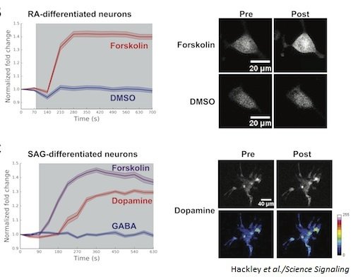
cAMPr: A genetically encoded single-wavelength fluorescent sensor for cyclic AMP. bit.ly/2BT8AJR @nyuschoolofmed @justinblau01 #cellimaging #BestOf2018

Labeling and Tracking Mitochondria with Photoactivation in Drosophila Embryos bio-protocol.org/e4347 #CellImaging #protocol @MBoCjournal @IISERPune

☑️ Gone are the days of manually tracing neurons from piles of Z-stacks. Explore automated segmentation using Aivia’s advanced machine learning capabilities. ☑️ Trial Aivia for free 👉 fcld.ly/aivia-lms-tw #microscopy #research #cellimaging #neuroscience

Labeling Endogenous Proteins Using #CRISPR-mediated Insertion of Exon (CRISPIE) bio-protocol.org/e4343 #CellImaging #cancer #protocol @eLife @OHSUNews

Nanolive at Institut Pasteur! 🎙️ Mathieu Fréchin shares insights on advanced cell characterization methods in his talk. Check out the collaborative work with @Virus_Immunity - Olivier Schwartz's group: bit.ly/3HFxWwZ #Biotech #CellImaging #Nanolive

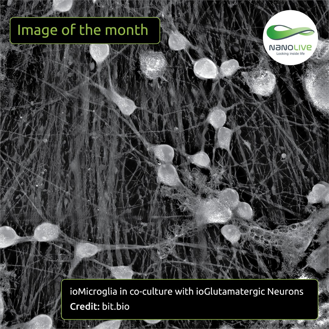
“Image of the Month”: Microglia cells patrol a neural network in vitro. Imaged during 3.5-hour using our 3D Cell Explorer 96focus. Thank you to @bitbio for sharing this beautiful image with us! 💫 bit.ly/4buVPW4 #ImageOfTheMonth #CellImaging #Nanolive

via #OSA_BOEx: Live-cell imaging of human spermatozoa using structured illumination microscopy ow.ly/U0KV30mClUa #Microscopy #CellImaging


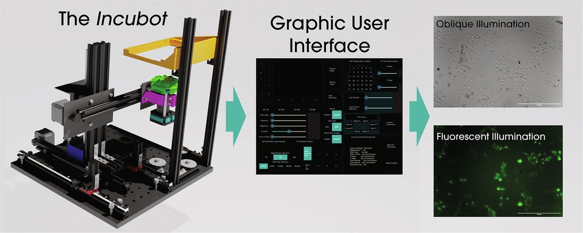
.@MercesGeorge, @PickeringLab, @ReynaudEmmanuel, et al. from @ucddublin published "The incubot: A 3D printer-based microscope for long-term live cell imaging within a tissue culture incubator". doi.org/10.1016/j.ohx.… #microscopy #opensource #cellimaging
🌟New series: "Image of the Month"! Label-free image of a mouse preadipocyte cell!🐭In this cell, organelles were captured at high resolution with Nanolive's 3D Cell Explorer-fluo, using our holotomographic tech. bit.ly/3NVtxJR #Imageofthemonth #Cellimaging #Nanolive


We are excited to share a collaboration starter kit between @gattaquant and @massphoton for your start into the world of #DNAPAINT. The bundle contains one PAINT 80R nanoruler slide and one MASSIVE-AB 1-PLEX labeling kit for #DNAPAINT #cellimaging. bit.ly/3dsh2S1

👩🔬🤝@Tomocubeinc and ibidi teams are excited to announce the joint release of the latest Tomocube HT Ready 96-well plate at the @ASCBiology #CellBio2023 Conference in Boston #collaboration #cellimaging #innovation #3Dcellimaging #technology #research #cellbiology #TechInnovation

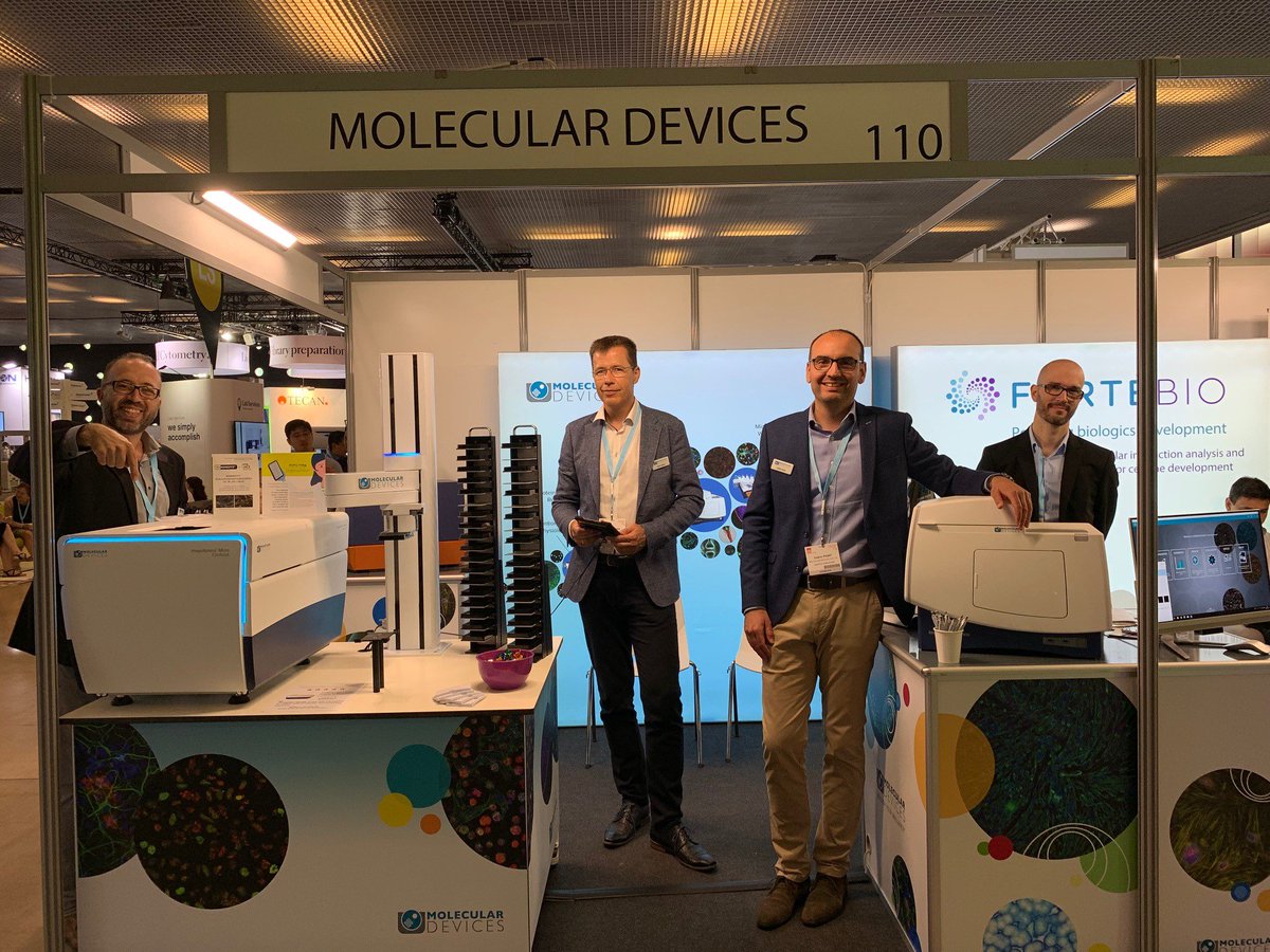
It’s the last day of #SLASEurope2019! We’re excited to present our range of #microplatereaders ,#cellimaging and #3Dimaging systems to accelerate your research. Visit us and @ForteBio at booths 101/110 and don't miss the poster on real time live cell assays #moleculardevices
🥼 Image of the month: Fluorescently labeled cancer cells with preadipocytes. Early apoptotic membrane blebbing visible after drug treatment. Explore more stunning images in our 2024 calendar: bit.ly/49DlU3B #ImageOfTheMonth #CellImaging #Nanolive


#MeetTheTeam Monday: Meet @emmajfong, a #CellImaging Staff Scientist in the @MumenthalerLab at the #EllisonInstitute! Emma earned her Ph.D. and MS in #ChemicalEngineering from @UCIrvine after receiving her BS in Chemical Engineering from @UCSanDiego.

Find out how a Canadian biotechnology company is harnessing cutting-edge real-time cell imaging to improve physiological relevance in intestinal organoids. Read the article: bit.ly/37P9CXD #organoids #cellimaging #livecellimaging #drugdevelopment #cellculture


A reversible #FluorescentProbe for real-time live #CellImaging and quantification of endogenous hydropolysulfides (Urano) @UTokyo_News_en doi.wiley.com/10.1002/anie.2…


Join this interesting Symposium S.19 'Brain in a dish – the past and future direction of hiPSC-derived cell models of neuropsychiatric disorders' at 8.30 CEST. #ECNP2022 @KittelSchneider #cellimaging

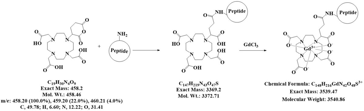
#mdpimolecules Imaging of Human Insulin Secreting Cells with Gd-DOTA-P88, a Paramagnetic Contrast Agent Targeting the Beta Cell Biomarker FXYD2γa mdpi.com/329908 #CellImaging @ULBruxelles
Something went wrong.
Something went wrong.
United States Trends
- 1. Cloudflare 209K posts
- 2. Gemini 3 23.3K posts
- 3. #AcousticPianoCollection 1,002 posts
- 4. Piggy 53K posts
- 5. Olivia Dean 3,929 posts
- 6. Taco Tuesday 14.4K posts
- 7. Saudi 112K posts
- 8. Anthropic 8,125 posts
- 9. Salman 29.4K posts
- 10. Good Tuesday 35.1K posts
- 11. #tuesdayvibe 3,063 posts
- 12. Sam Leavitt N/A
- 13. #ONEPIECE1166 4,275 posts
- 14. CAIR 23K posts
- 15. JUST ANNOUNCED 26.2K posts
- 16. jeonghan 93.1K posts
- 17. Gary Sinise 6,476 posts
- 18. Garling 2,236 posts
- 19. Passan N/A
- 20. Brian Walshe N/A