#micrographs search results
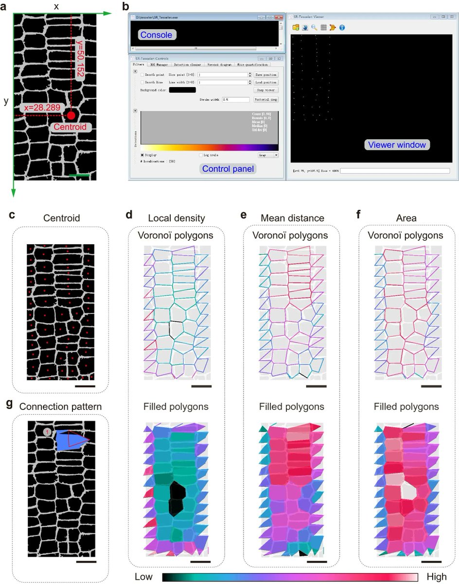
High-efficiency procedure to characterize, segment, and quantify complex multicellularity in raw micrographs in plants plantmethods.biomedcentral.com/articles/10.11… #plantsci #raw #micrographs ♻️


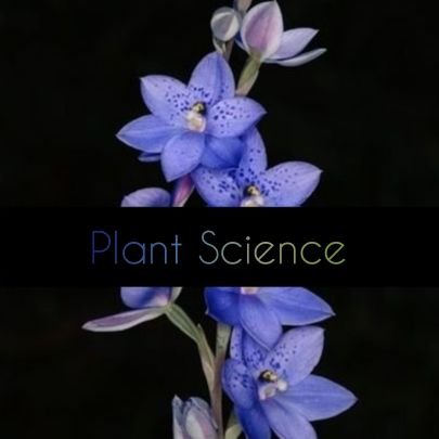
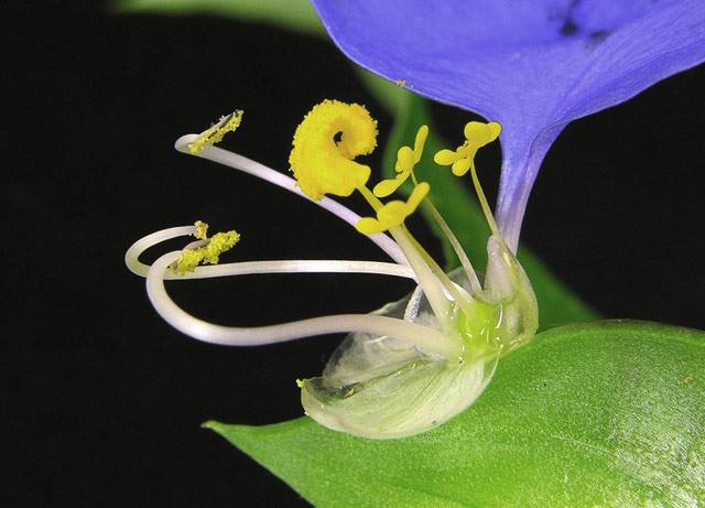
Happy #NationalWildflowerWeek! A few of our favorite flower #micrographs from the archives: bit.ly/1h0lh8h

cold work micrographs Powerpoint Templates - ppthunter.com/cold-work-micr… #cold #work #micrographs
The Beautiful Secrets of Tears, Revealed By a Microscope gizmodo.com/the-beautiful-… Thank you @nattilak ! #micrographs #microscope #tears

We are accepting images for the 2015 RMS Calendar. Submissions to [email protected] by Nov 14th #micrographs #microscopy


Did you ever find a surprise in your SEM image? Our applications scientist did! This dragon appeared in a sample of Dragonite he was imaging with the SEM. #halloween #micrographs #happyhalloween #letsgetspooky


From your #micrographs to #dronegrams to #astrophotos, our #lithophanes can make them awesome! #3dprinting @lulzbot3d #makersgonnamake


Light #micrographs, #3D tumour #tomography, and optical tissue clearing images reveal the "The colour of #cancer" - dailym.ai/2e7vKnp
What are some good #books that collect #electron #micrographs of #cardiacmuscle, especially all the amazing work from the 70's and 80's? #Microscopy, #Cardiology peeps, please chime in.
We dug up a theme for this week! Snow! It's coming our way to New England. Enjoy these snow-related Scanning Electron Microscope images - with descriptions of what they really are (except one really is a snowflake). #micrographs #SEM #microscopy #bombogenesis #snow #snowday

One of the most stunning #micrographs I have ever seen. Science can be truly beautiful, don't you think? fb.me/4QiVoIDX7
We are now welcoming #micrographs to the 2017 RMS Scientific Imaging Competition rms.org.uk/discover-engag…

A gallery of #micrographs turns cells into visual art on #DiscoveryUnbound via @Wired 🔬 bit.ly/1THTAQf

Check out the new web site for the Australian #ceramics Society that features @TESCAN #SEM #micrographs #microscope austceram.com


False-colored transmission electron micrograph of an aortic smooth muscle cell from a mouse with progeria. Submitted by: Thomas Weston, University of California Los Angeles, Los Angeles, CA #MicroscopyMonday #Micrographs


Great #micrographs by the @UMBC Keith R. Porter Imaging Facility manager, @nerd__candy !!! @UMBCBiology @UMBCCNMS @HHMIBilly #SEAPHAGES

At 3/4 images (n=45) here are Siphos MarshalMathers (Venice, Italy) and Mouse (Silver Spring), and Podos Brittakah & GreenThumb from @UMBC
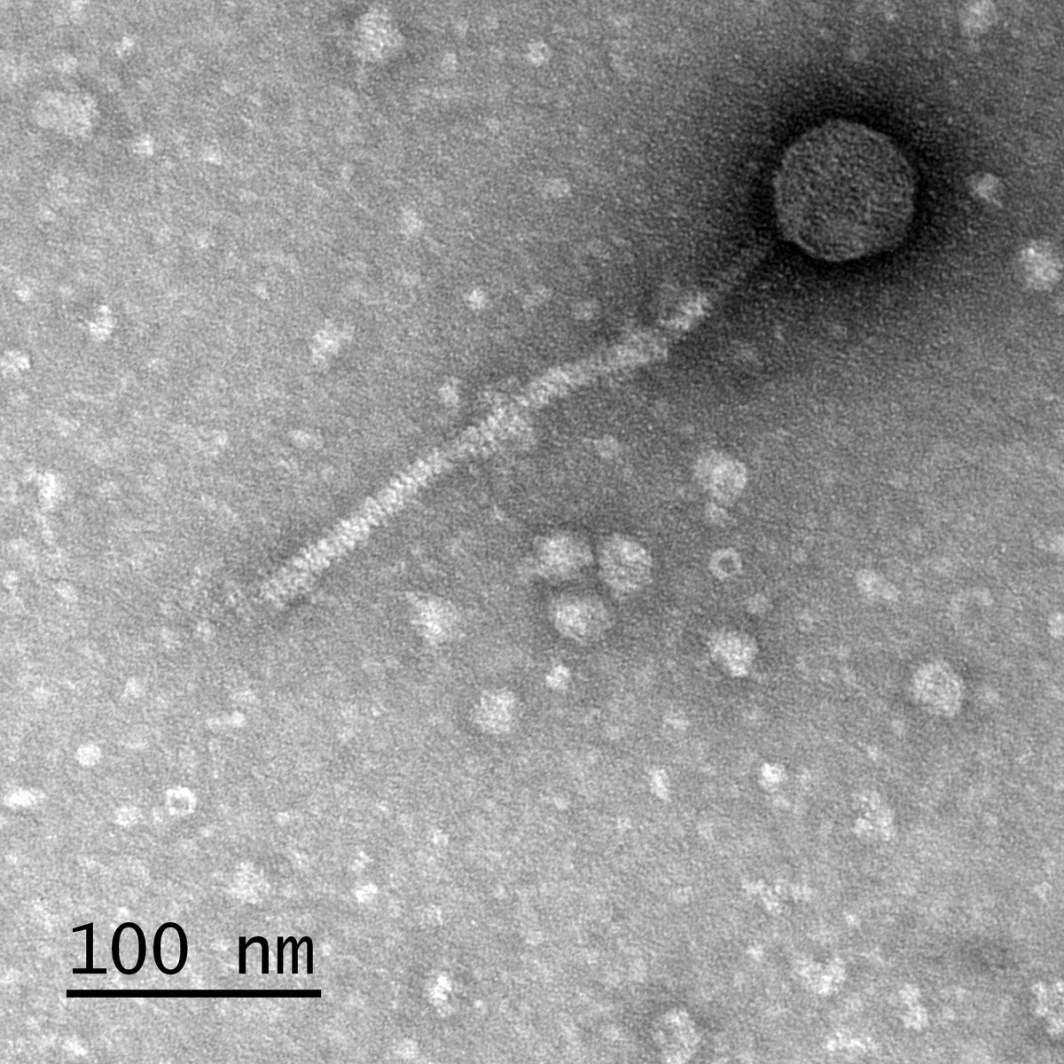
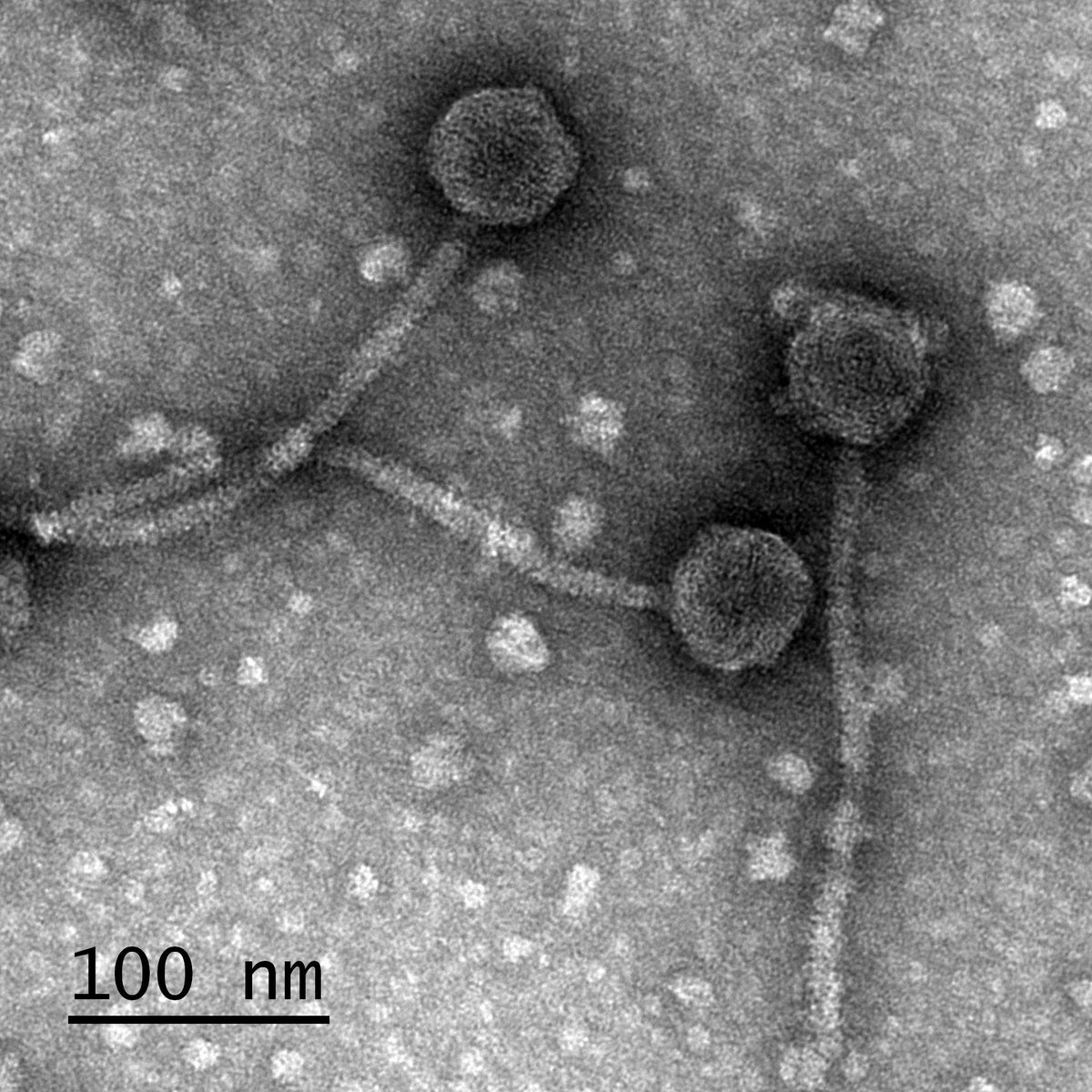

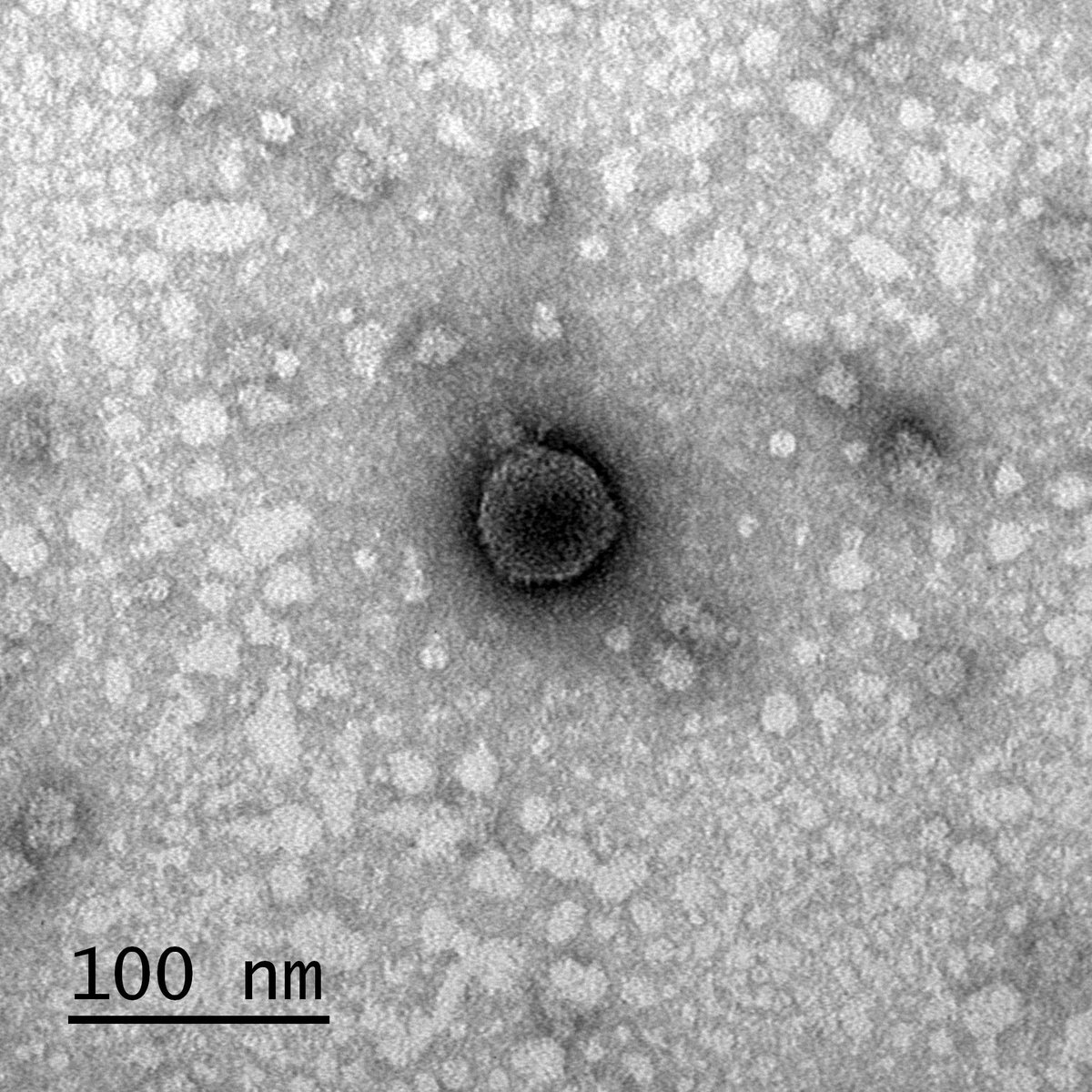

Photo: Close-Up Cuisine: Terra Cibus No. 4 Fortune Cookie tmblr.co/ZfxagxEDGUvi #tumblr #micrographs
I’ll try it on little grape grafts #MicroGraphs

Tomato Grafting Clips on Amazon ✊✊ You can use grafting tape but I prefer to use clips and a ziplock bag!
False-colored transmission electron micrograph of an aortic smooth muscle cell from a mouse with progeria. Submitted by: Thomas Weston, University of California Los Angeles, Los Angeles, CA #MicroscopyMonday #Micrographs

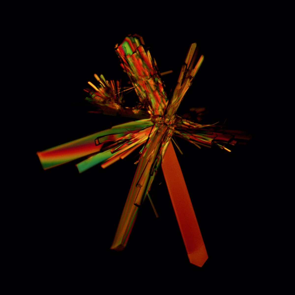
This #micrographs that show yellowish-brown #mica #inclusions (biotite-phlogopite series) in an #emerald from #Zambia (#Kafubu deposit). This micrograph is taken in polarised light (crossed polarisers), which induces interference colours visible in the large central #crystal.


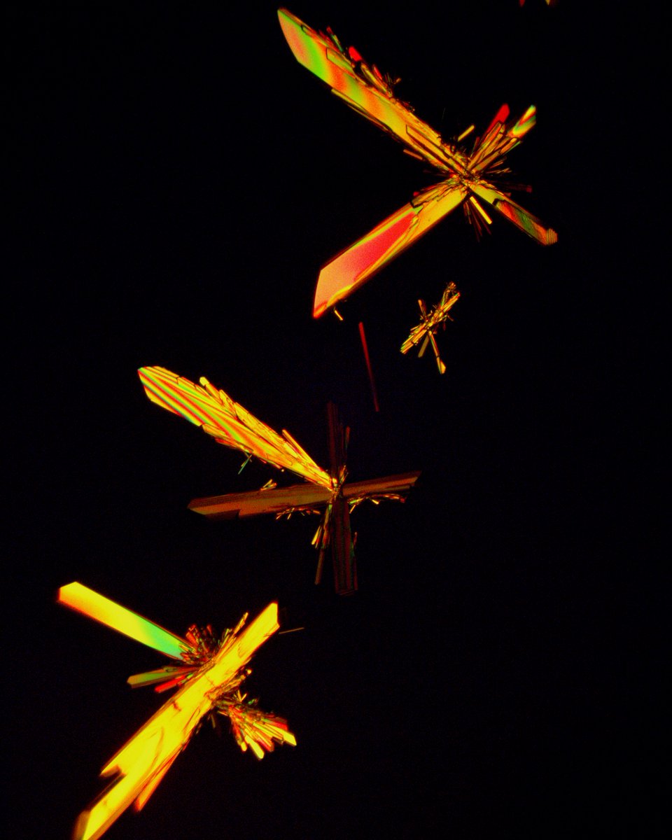
Two-amino-acid Snow. Two amino acids, glutamine and alanine, recrystallized from solution onto a microscope slide into discrete crystal formations. One reminded me of a drum major. Another grouping took on an entomological vibe. #scicomm #micrographs
Day 2 #Coursera, image processing, I need the #MatLab refresher, turns out I have access as University staff. It's all coming back, none of it is #micrographs, but it's all the same terms. So, tonight, MatLab, tomorrow back to Coursera. #100DaysOfCode #ImageProcessing
Did you ever find a surprise in your SEM image? Our applications scientist did! This dragon appeared in a sample of Dragonite he was imaging with the SEM. #halloween #micrographs #happyhalloween #letsgetspooky

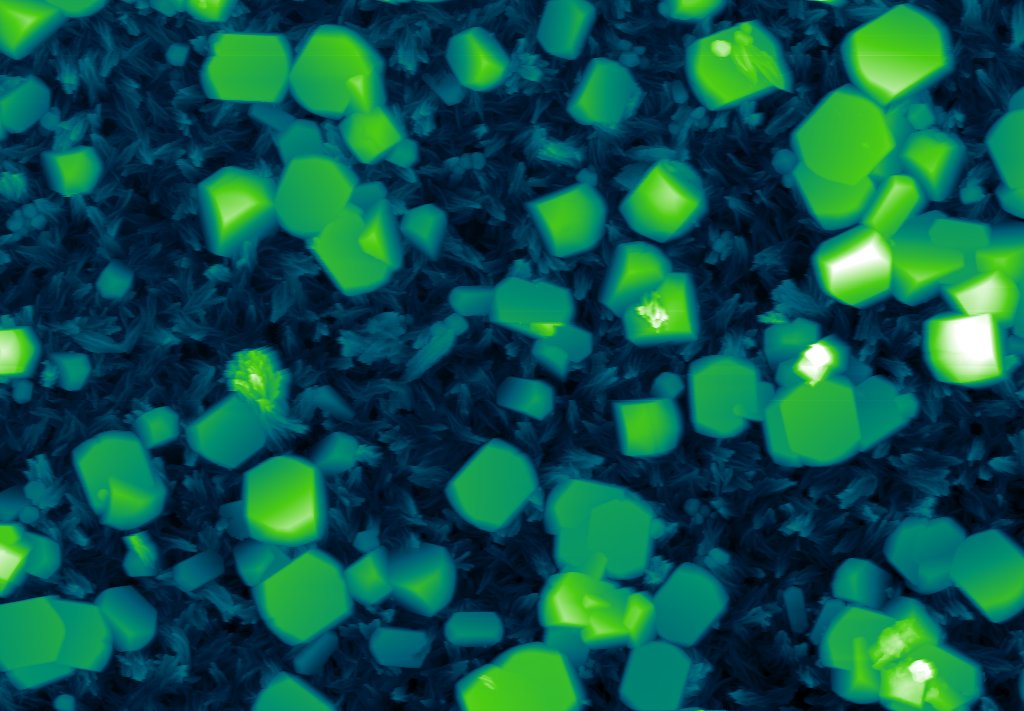
phys.org/news/2022-09-f… #micrographs of alkaline-earth chalcogenide (#AeCh) #nanocrystals
phys.org
Fundamental research improves understanding of new optical materials
Research into the synthesis of new materials could lead to more sustainable and environmentally friendly items such as solar panels and light emitting diodes (LEDs). Scientists from Ames National...
What are some good #books that collect #electron #micrographs of #cardiacmuscle, especially all the amazing work from the 70's and 80's? #Microscopy, #Cardiology peeps, please chime in.

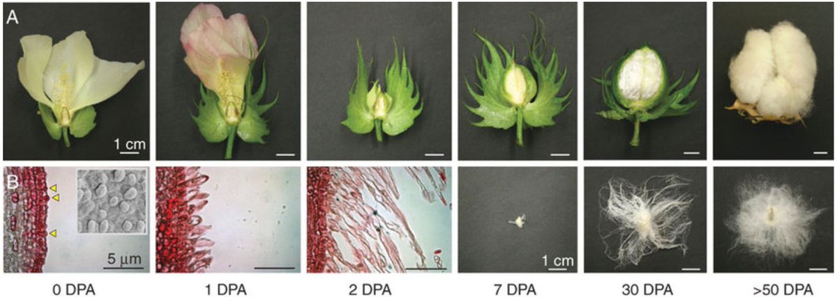
It’s #cotton #fact Friday! The images & #micrographs below show the cotton development process 👇 When the cotton flower turns pink, it has self-pollinated and the cells on the outside of the #seed start to elongate & mature 🌸 #AgBiTech 📷 Andrew W Woodward, ResearchGate

We like to share overall industry news as well as insights into unique projects in the space. Micrographs seeks to unlock the biggest barrier to blockchain adoption globally… @micrographs_io #micrographs #Blockchain #Crypto #CryptoNews thelatestblock.com/what-is-the-bi…
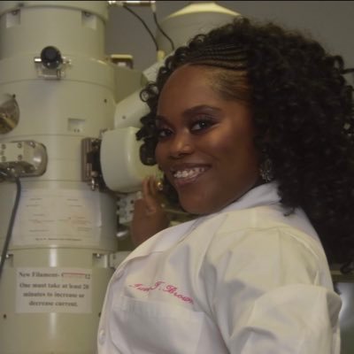
And yes, that’s my beautiful nail we’re putting in focus on a @LeicaMicro 🔬! Because that’s the fun and power in microscopy !!! 😃😍 (and because we could, so why not ?) • What’s the best thing you’ve imaged under a microscope? #microscopetales of #micrographs

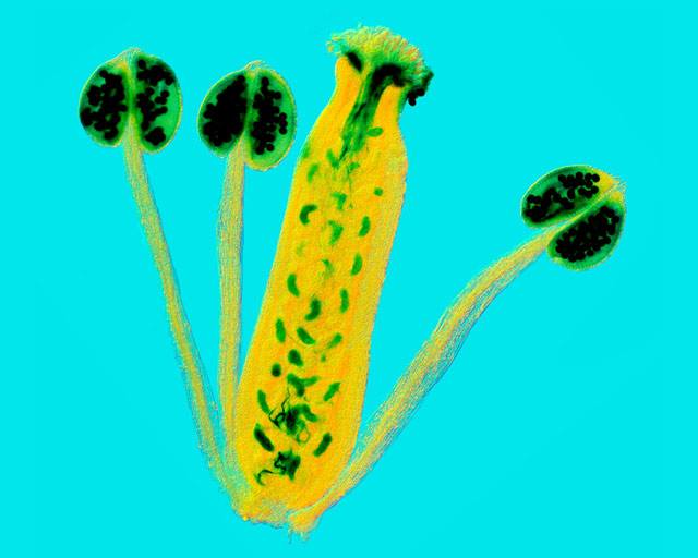
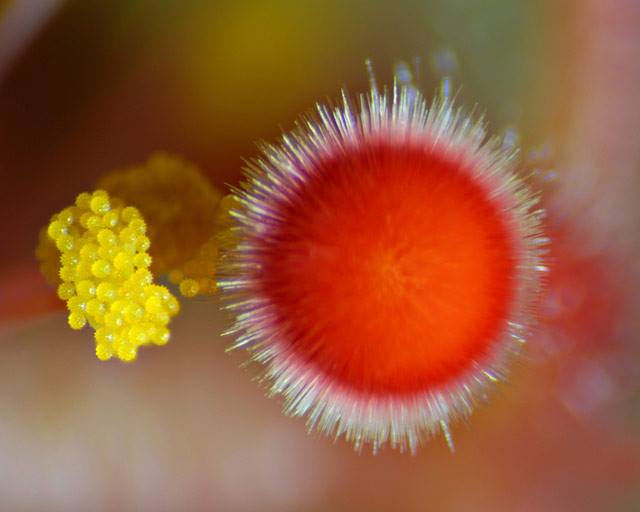
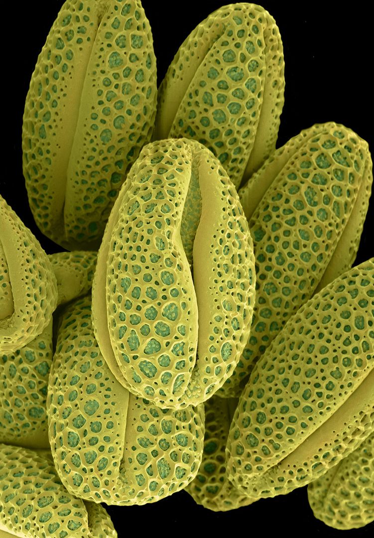
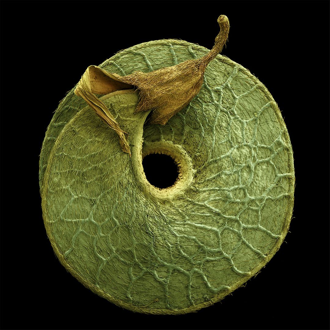
#Science #Art: colored #micrographs magnify #pollen seeds, plant cells, and leaf structures in photographs by Rob Kesseler thisiscolossal.com/2019/12/rob-ke… via @Colossal


High-efficiency procedure to characterize, segment, and quantify complex multicellularity in raw micrographs in plants plantmethods.biomedcentral.com/articles/10.11… #plantsci #raw #micrographs ♻️
Our Enhancing your Image Course is taking place today at @JohnInnesCentre We can't wait to see the beautiful #micrographs that are created

We are now welcoming #micrographs to the 2017 RMS Scientific Imaging Competition rms.org.uk/discover-engag…
We're inviting #micrographs to this year's Scientific Imaging Competition. Get your best image noticed! rms.org.uk/discover-engag… @benaviss


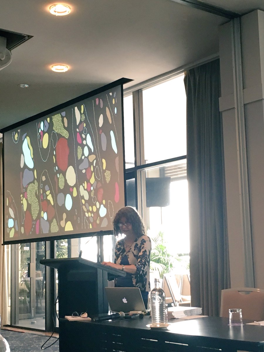
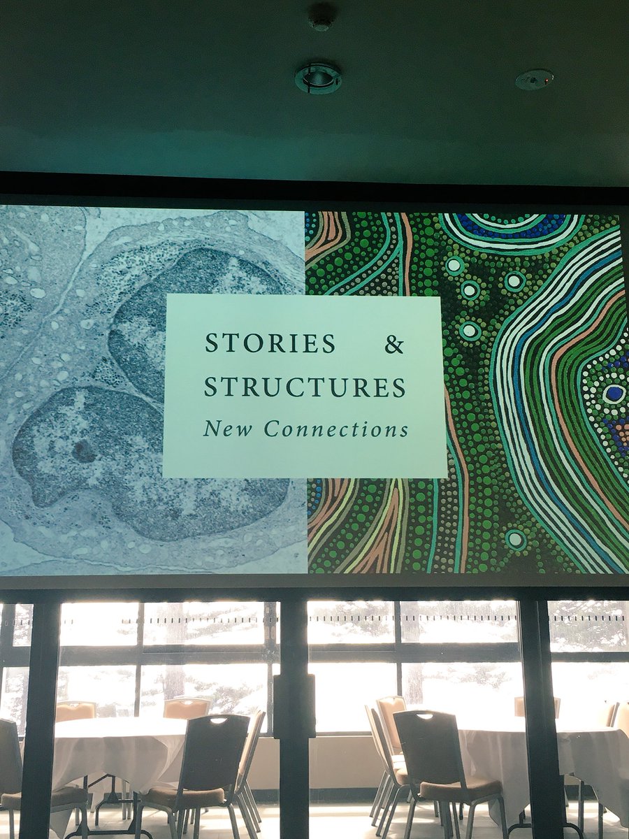
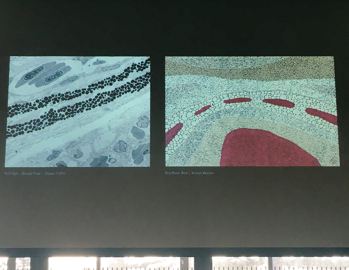
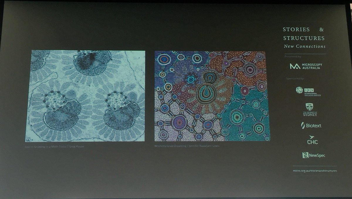
Starting Day 2 of #FutureMicrobiome2018: Jenny Whiting speaks about a wonderful exhibition of #micrographs and Aboriginal Australian #art she has curated. Check it out here: micro.org.au/storiesandstru…

Coloured #Micrographs Magnify #Pollen Seeds, Plant Cells, and Leaf Structures in Photographs by Rob Kesseler ow.ly/tOgb30pZUbg
Colored #Micrographs Magnify #Pollen #Seeds, #Plant #Cells, and #Leaf Structures in #Photographs by #RobKesseler thisiscolossal.com/2019/12/rob-ke… #macrophotography #microscopy


The ultrastructure of infectious L-type bovine spongiform #encephalopathy prions constrains molecular models: ow.ly/ChWF50F4kbT Additional raw #data (electron #micrographs) and 3D reconstructions are deposited in figshare: ow.ly/NK0J50F4kba ow.ly/T9Xh50F4kbb


Coloured #Micrographs Magnify #Pollen Seeds, Plant Cells, and Leaf Structures in Photographs by Rob Kesseler ift.tt/2PshaaE ift.tt/35s7Adl

From your #micrographs to #dronegrams to #astrophotos, our #lithophanes can make them awesome! #3dprinting @lulzbot3d #makersgonnamake

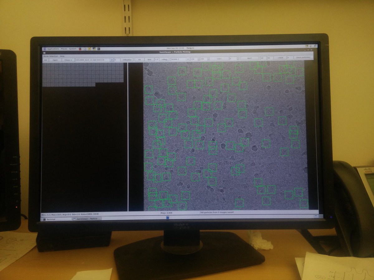
#particlepicking on these icy #micrographs.. Not so cool.. #CryoEM #structuralbiology #pdbem #onedayi'llhavehighresolution #sigh

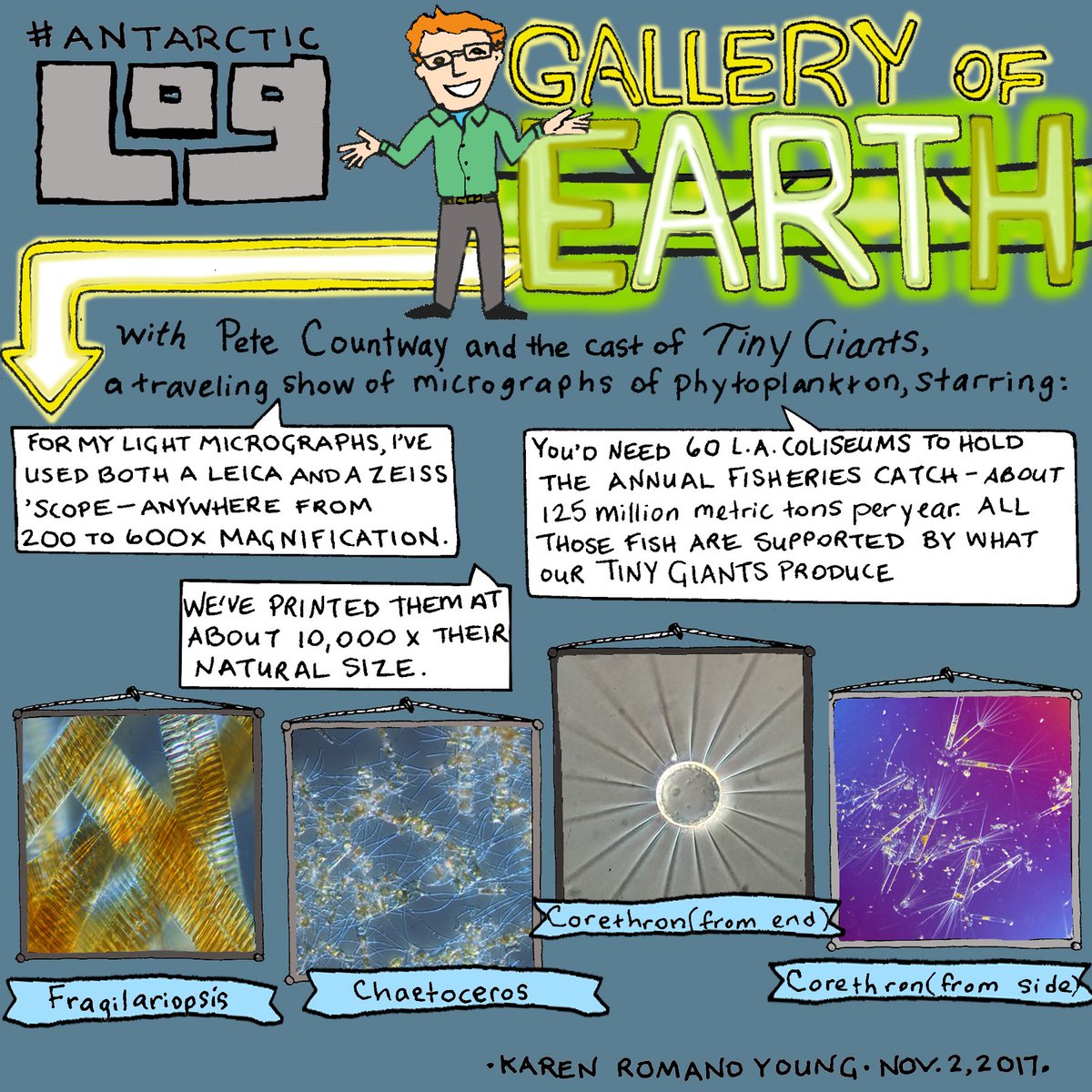
Little things... #AntarcticLog focuses on #micrographs from @BigelowLab 's #TinyGiants exhibit. #Scienceandart #STEAM #STEM #sciencecomics
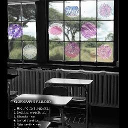
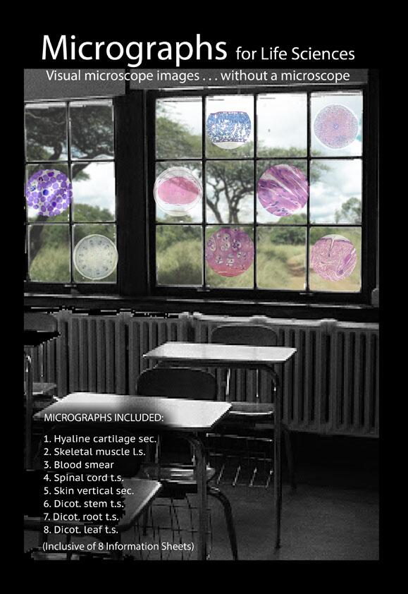
@dragonjones #Micrographs for schools without #microscopes . These are use in #rural #poor #poverty #owninvention
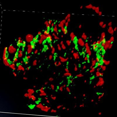
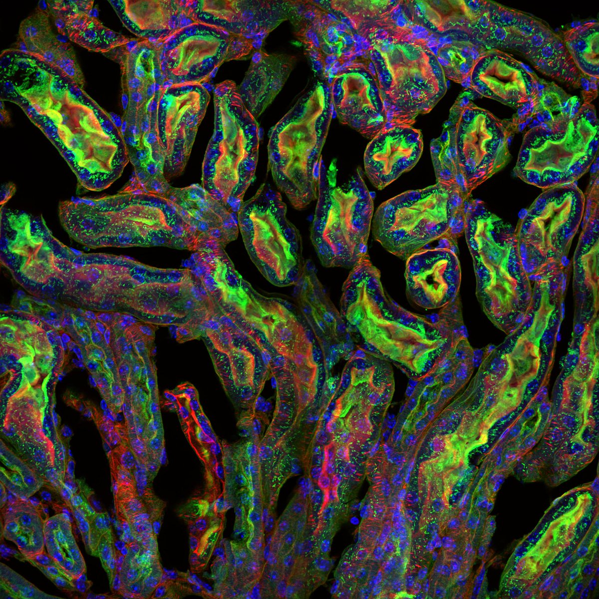
Check out these #micrographs of a mouse kidney section captured on the SpinSR super resolution microscope.


Amazing Micrographs Show What Cells Really… dlvr.it/CtnK68 #cellbiology #gallery #micrographs #Science

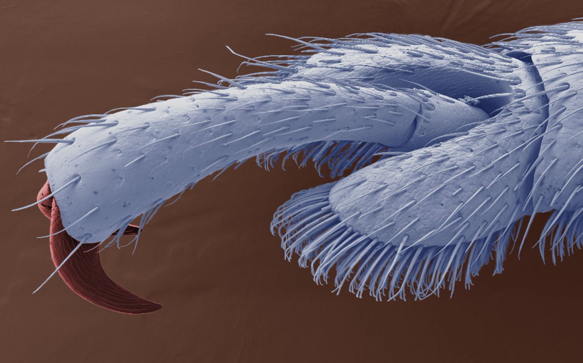
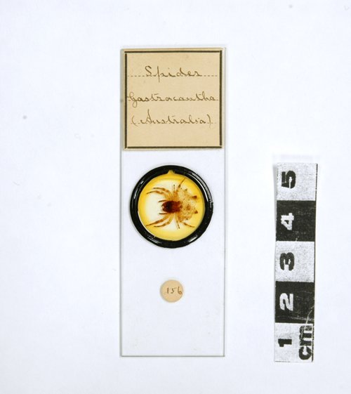
I love the #micrographs that were taken from #microscope slides in the C.R. Percival #Collection (1895-1954) at the @SciTechMuseum. You can see a sample of the images online just like this orb-weaver #spider at: ingeniumcanada.org/scitech/whats-… #heritageMW #MuseumWeek #science #biology
Happy #NationalWildflowerWeek! A few of our favorite flower #micrographs from the archives: bit.ly/1h0lh8h

smashing book! Found in the kids section of our public library. All SEM images #micrographs #morphology #insects
Something went wrong.
Something went wrong.
United States Trends
- 1. Iran N/A
- 2. TACO N/A
- 3. #WWENXT N/A
- 4. Amed Rosario N/A
- 5. Strait N/A
- 6. #DaredevilBornAgain N/A
- 7. #HighPotential N/A
- 8. Ceasefire N/A
- 9. Vrabel N/A
- 10. Pakistan N/A
- 11. Art of the Deal N/A
- 12. #ATOBTTR N/A
- 13. Lamelo N/A
- 14. Mark Leiter Jr N/A
- 15. Russini N/A
- 16. Taj Bradley N/A
- 17. Sion James N/A
- 18. Mythos N/A
- 19. Sandy N/A
- 20. Wisconsin Supreme Court N/A