#micrographs search results
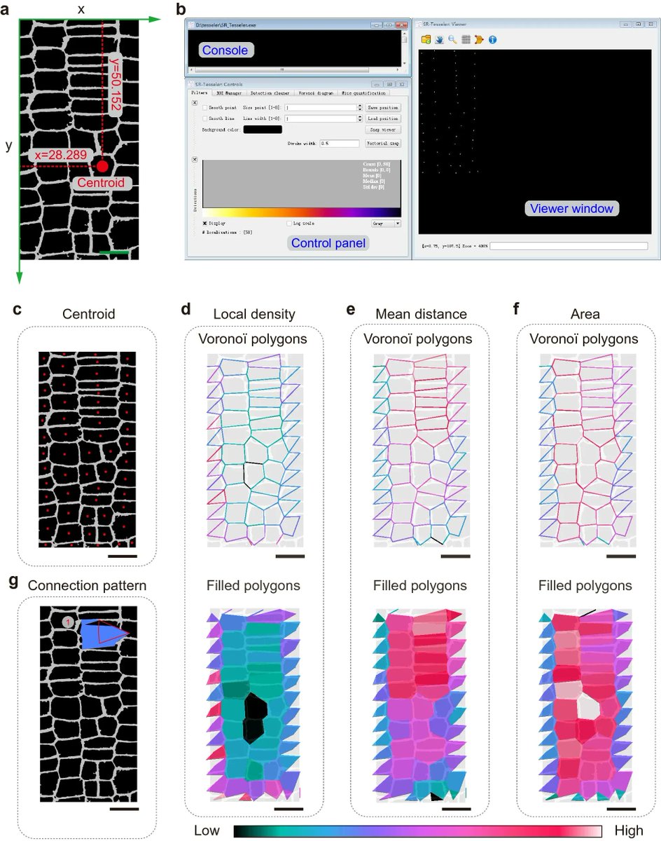
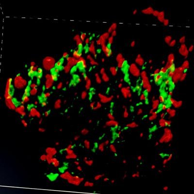
High-efficiency procedure to characterize, segment, and quantify complex multicellularity in raw micrographs in plants plantmethods.biomedcentral.com/articles/10.11… #plantsci #raw #micrographs ♻️


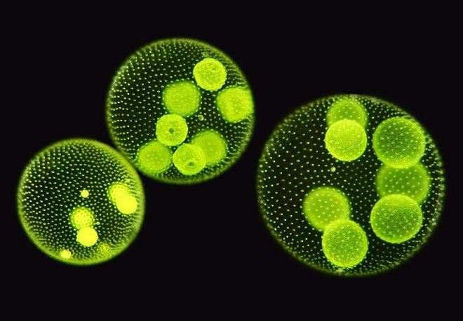
Amazing #Micrographs Show What Cells Really Look Like goo.gl/fb/Swb2r6 #cellbiology #gallery #science

Light #micrographs of an Esophageal #Cancer forming Epithelial #Cell Nests. tmblr.co/ZK5Bsr1FrOF2o #stock #medical #images

One of the most stunning #micrographs I have ever seen. Science can be truly beautiful, don't you think? fb.me/4QiVoIDX7
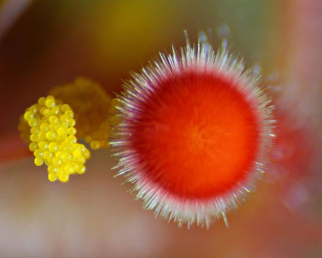
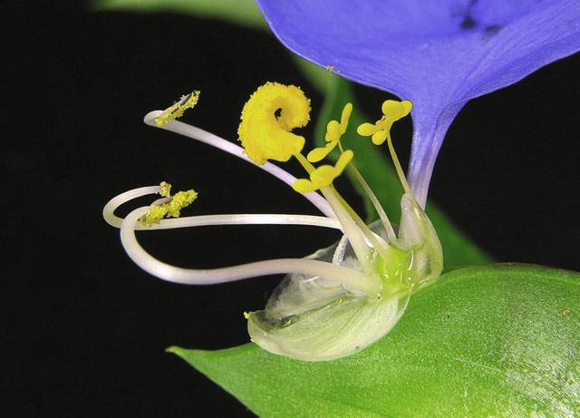
Happy #NationalWildflowerWeek! A few of our favorite flower #micrographs from the archives: bit.ly/1h0lh8h

are you trashing 90% of your TEM pictures?? too bad bit.ly/WY7HlZ #micrographs

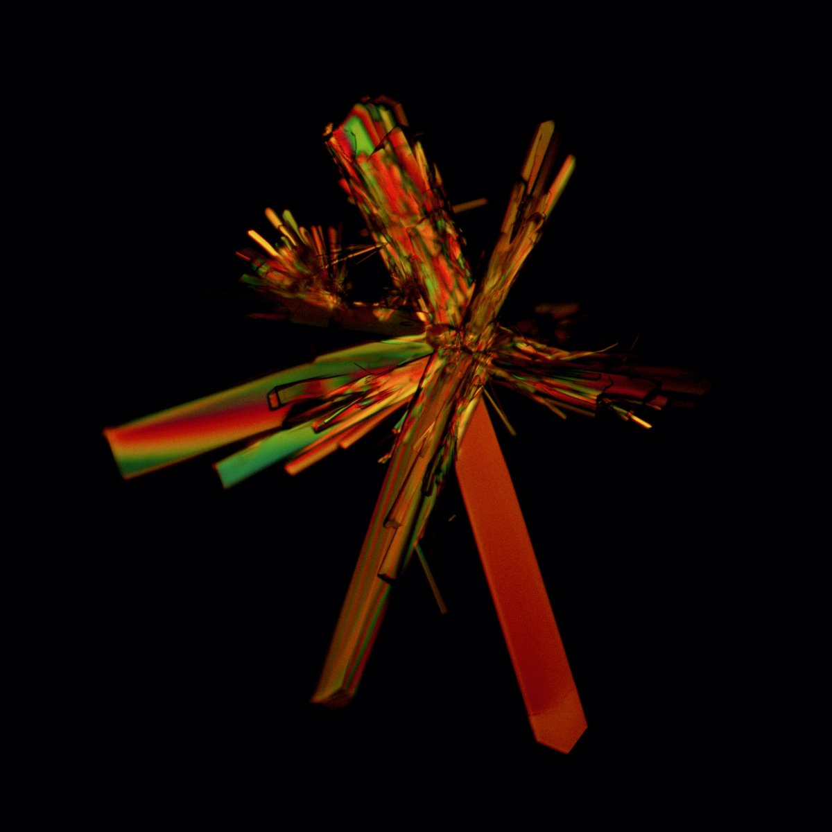
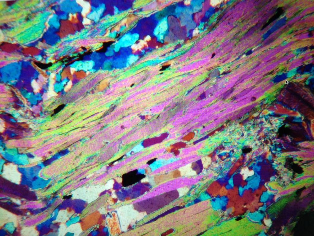
This #micrographs that show yellowish-brown #mica #inclusions (biotite-phlogopite series) in an #emerald from #Zambia (#Kafubu deposit). This micrograph is taken in polarised light (crossed polarisers), which induces interference colours visible in the large central #crystal.


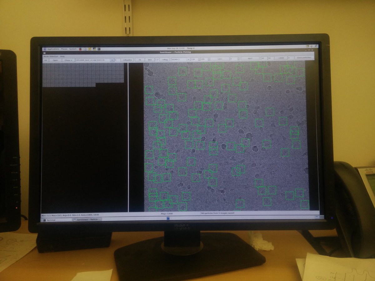
#particlepicking on these icy #micrographs.. Not so cool.. #CryoEM #structuralbiology #pdbem #onedayi'llhavehighresolution #sigh
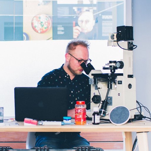
From parts to pictures (#micrographs)!
@GeoSciTweeps We need to talk this week. I just got my ISI Super IIIA running, it started as a truckload of parts and now I am getting this.


The ultrastructure of infectious L-type bovine spongiform #encephalopathy prions constrains molecular models: ow.ly/ChWF50F4kbT Additional raw #data (electron #micrographs) and 3D reconstructions are deposited in figshare: ow.ly/NK0J50F4kba ow.ly/T9Xh50F4kbb


Amazing Micrographs Show What Cells Really… dlvr.it/CtnK68 #cellbiology #gallery #micrographs #Science
The Beautiful Secrets of Tears, Revealed By a Microscope gizmodo.com/the-beautiful-… Thank you @nattilak ! #micrographs #microscope #tears

I've been staring at #histology #micrographs too long today, they're starting to stare back!! :O #spleen #resinsection @Chapman_Histo @VMDatabase
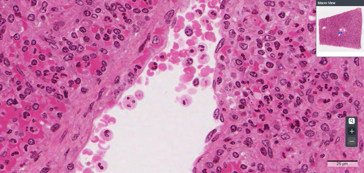

Photo: Close-Up Cuisine: Terra Cibus No. 4 Fortune Cookie tmblr.co/ZfxagxEDGUvi #tumblr #micrographs
Just a case of #nerves. See our beautiful #micrographs of human nerve fibers. (bit.ly/16mhDOK) #stockimages
There are fewer things in life that are finer than #microscopy themed calendars. How else are you supposed to track your resolutions? #afm #micrographs #free

"Nano is Art - Park AFM Calendars" Available Free To all our followers in the US. Send an email to [email protected] with your address to get your Park AFM Calendar. #Nanoart #ParkSystems #nanocalendar2021


I’ll try it on little grape grafts #MicroGraphs

Tomato Grafting Clips on Amazon ✊✊ You can use grafting tape but I prefer to use clips and a ziplock bag!

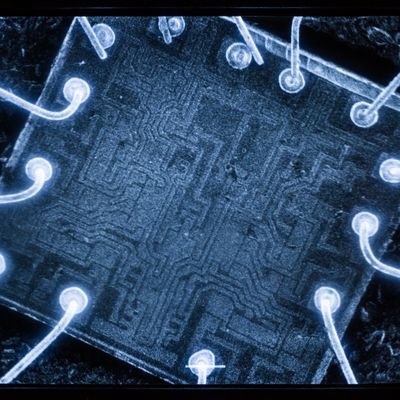
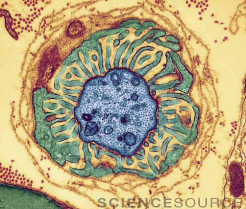
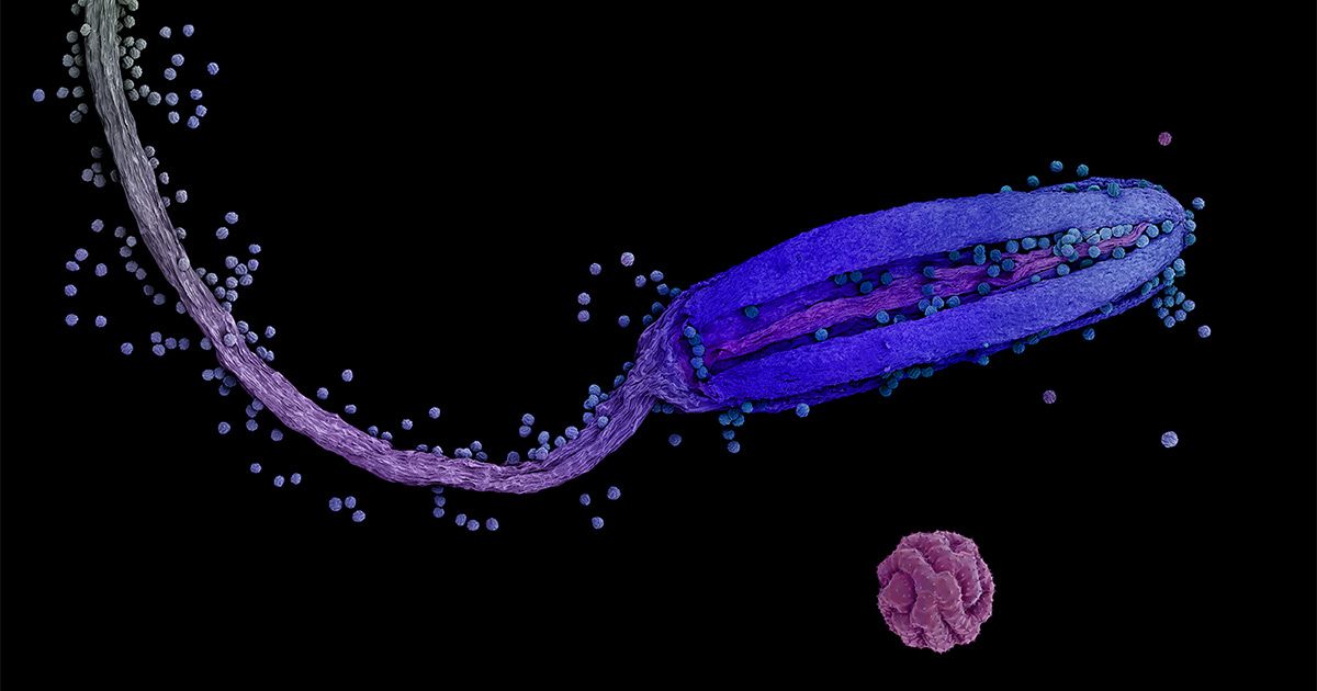
False-colored transmission electron micrograph of an aortic smooth muscle cell from a mouse with progeria. Submitted by: Thomas Weston, University of California Los Angeles, Los Angeles, CA #MicroscopyMonday #Micrographs

This #micrographs that show yellowish-brown #mica #inclusions (biotite-phlogopite series) in an #emerald from #Zambia (#Kafubu deposit). This micrograph is taken in polarised light (crossed polarisers), which induces interference colours visible in the large central #crystal.

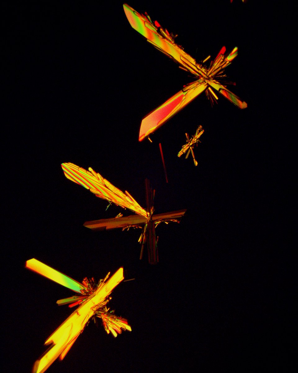
Two-amino-acid Snow. Two amino acids, glutamine and alanine, recrystallized from solution onto a microscope slide into discrete crystal formations. One reminded me of a drum major. Another grouping took on an entomological vibe. #scicomm #micrographs
Day 2 #Coursera, image processing, I need the #MatLab refresher, turns out I have access as University staff. It's all coming back, none of it is #micrographs, but it's all the same terms. So, tonight, MatLab, tomorrow back to Coursera. #100DaysOfCode #ImageProcessing

Did you ever find a surprise in your SEM image? Our applications scientist did! This dragon appeared in a sample of Dragonite he was imaging with the SEM. #halloween #micrographs #happyhalloween #letsgetspooky


phys.org/news/2022-09-f… #micrographs of alkaline-earth chalcogenide (#AeCh) #nanocrystals

What are some good #books that collect #electron #micrographs of #cardiacmuscle, especially all the amazing work from the 70's and 80's? #Microscopy, #Cardiology peeps, please chime in.

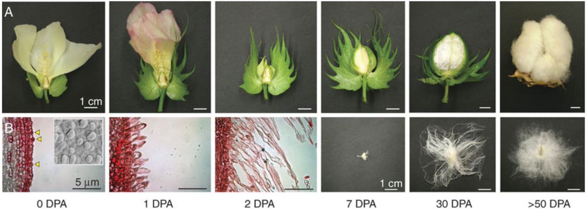
It’s #cotton #fact Friday! The images & #micrographs below show the cotton development process 👇 When the cotton flower turns pink, it has self-pollinated and the cells on the outside of the #seed start to elongate & mature 🌸 #AgBiTech 📷 Andrew W Woodward, ResearchGate

We like to share overall industry news as well as insights into unique projects in the space. Micrographs seeks to unlock the biggest barrier to blockchain adoption globally… @micrographs_io #micrographs #Blockchain #Crypto #CryptoNews thelatestblock.com/what-is-the-bi…
And yes, that’s my beautiful nail we’re putting in focus on a @LeicaMicro 🔬! Because that’s the fun and power in microscopy !!! 😃😍 (and because we could, so why not ?) • What’s the best thing you’ve imaged under a microscope? #microscopetales of #micrographs

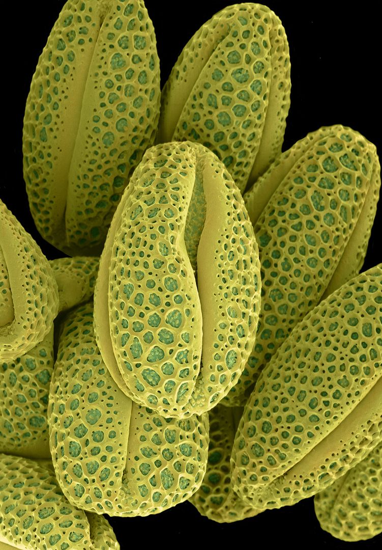
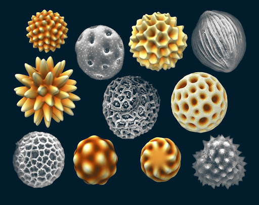
#Science #Art: colored #micrographs magnify #pollen seeds, plant cells, and leaf structures in photographs by Rob Kesseler thisiscolossal.com/2019/12/rob-ke… via @Colossal


High-efficiency procedure to characterize, segment, and quantify complex multicellularity in raw micrographs in plants plantmethods.biomedcentral.com/articles/10.11… #plantsci #raw #micrographs ♻️

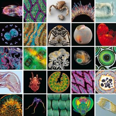
Clever human stuff. #micrographs #science #stem See the big winners of Nikon’s micro-photo competition
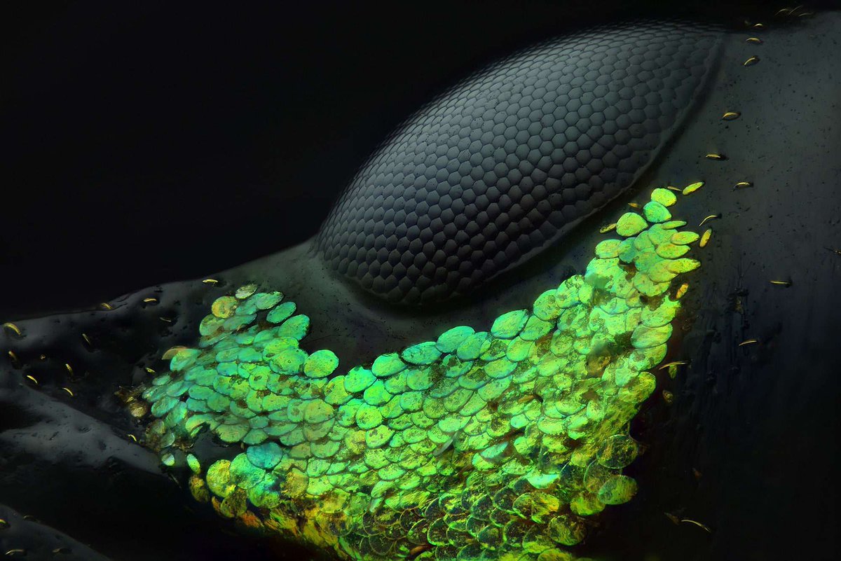

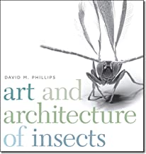
smashing book! Found in the kids section of our public library. All SEM images #micrographs #morphology #insects
Amazing Micrographs Show What Cells Really… dlvr.it/CtnK68 #cellbiology #gallery #micrographs #Science


#particlepicking on these icy #micrographs.. Not so cool.. #CryoEM #structuralbiology #pdbem #onedayi'llhavehighresolution #sigh
We're on instagram! Tag us in your #micrographs, microscope selfies or anything #microscopy! instagram.com/microscopy_soc/

From your #micrographs to #dronegrams to #astrophotos, our #lithophanes can make them awesome! #3dprinting @lulzbot3d #makersgonnamake

Happy #NationalWildflowerWeek! A few of our favorite flower #micrographs from the archives: bit.ly/1h0lh8h
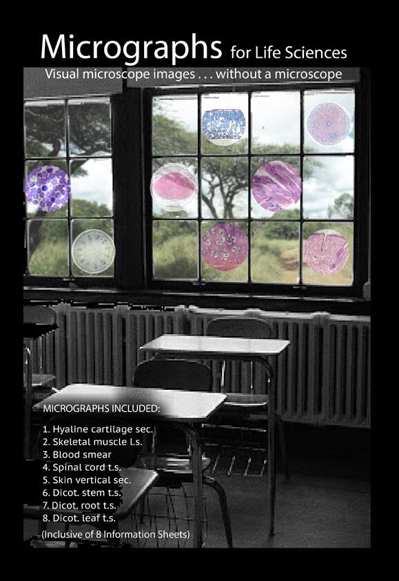
@richardbranson #Micrographs for schools without #microscopes . Please RT #owninvention rural Africa rural Americas


This #micrographs that show yellowish-brown #mica #inclusions (biotite-phlogopite series) in an #emerald from #Zambia (#Kafubu deposit). This micrograph is taken in polarised light (crossed polarisers), which induces interference colours visible in the large central #crystal.

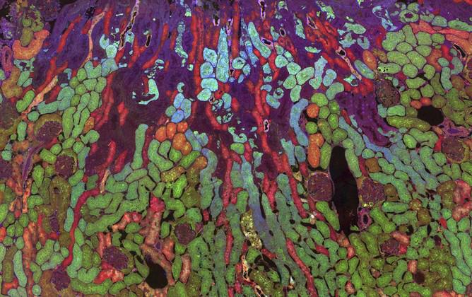
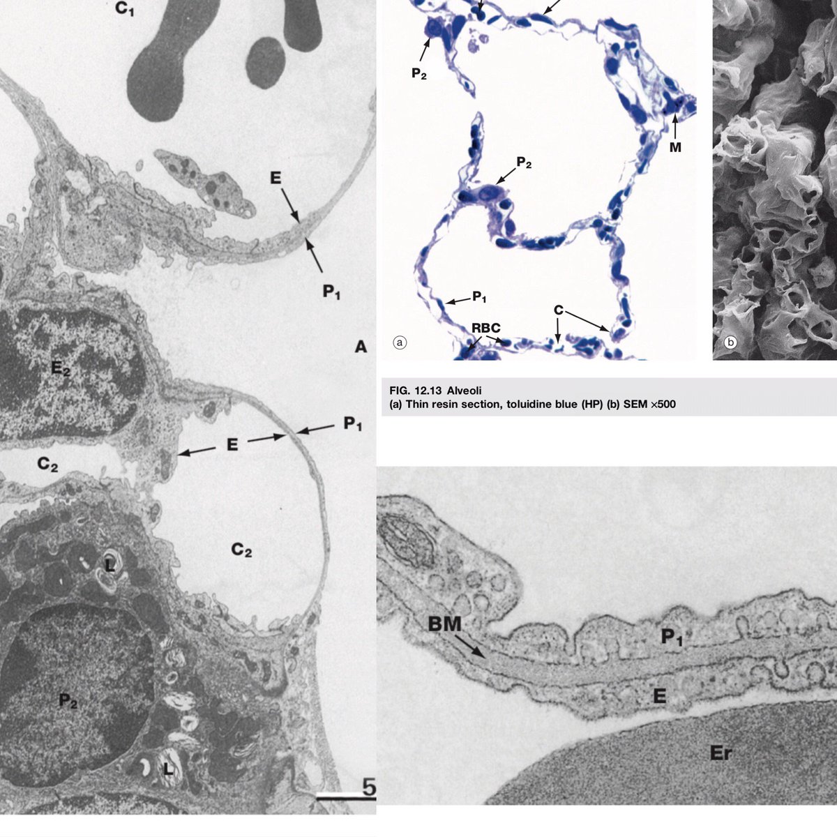
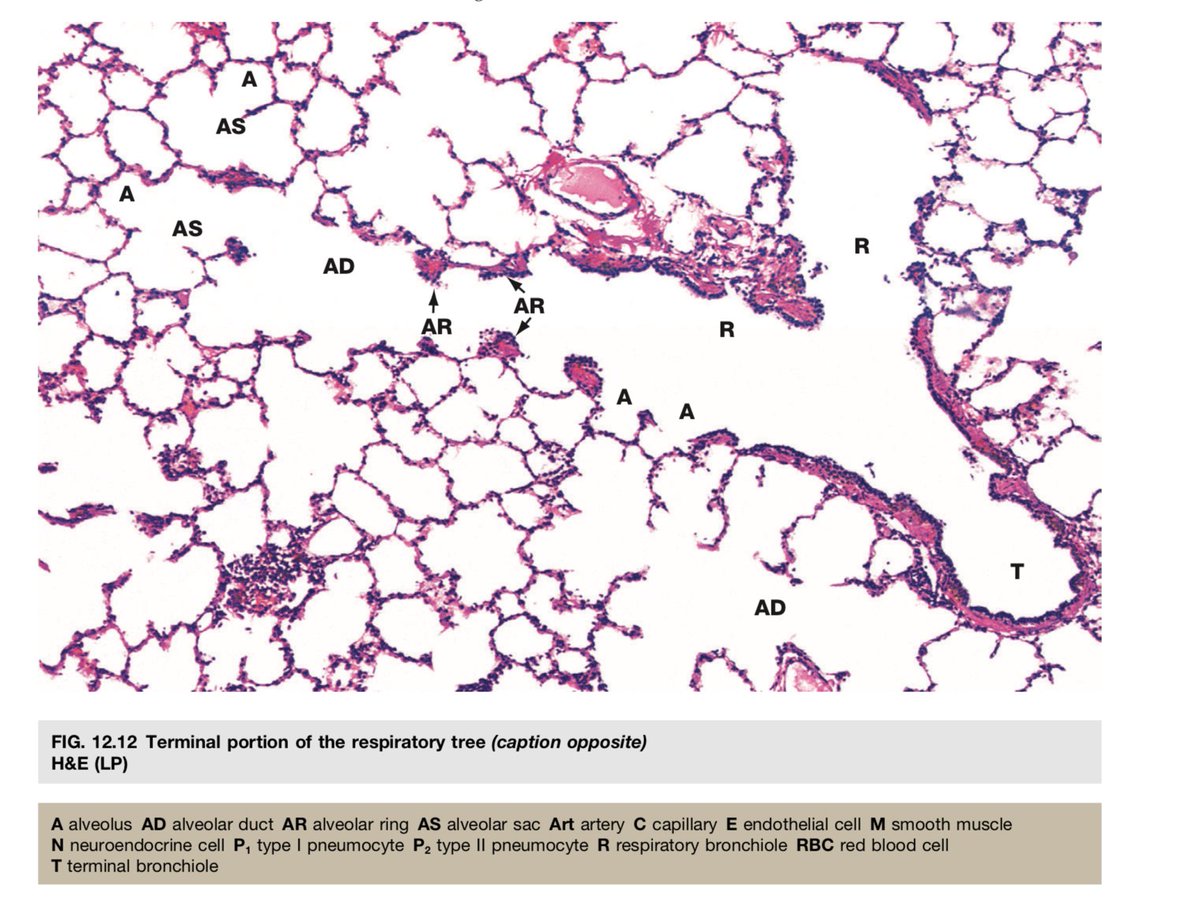
🔬Micro time! These are #lung #micrographs - can you identify alveoli, RBCs, and capillaries? Type I pneumocytes are large, flat, and are the barrier for gas exchange. Type II are cuboidal, produce surfactant, and are stem cells. Photos from Wheater’s Functional #Histology.
#Micrographs allows #developers to easily export popular #blockchain services and smart contracts as REST APIs. These APIs can also be used with cloud-native architectures (Kubernetes, Docker), or to develop entire #business apps within our ecosystem. 👉🏼micrographs.io


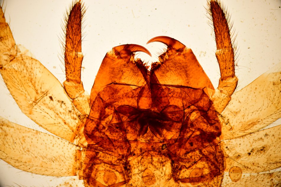
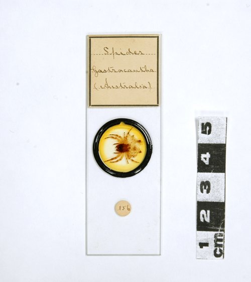
I love the #micrographs that were taken from #microscope slides in the C.R. Percival #Collection (1895-1954) at the @SciTechMuseum. You can see a sample of the images online just like this orb-weaver #spider at: ingeniumcanada.org/scitech/whats-… #heritageMW #MuseumWeek #science #biology
Two-amino-acid Snow. Two amino acids, glutamine and alanine, recrystallized from solution onto a microscope slide into discrete crystal formations. One reminded me of a drum major. Another grouping took on an entomological vibe. #scicomm #micrographs
“The Scream” by Munch with liposomes on the nanoscale (and many other funny #TEM #micrographs) bit.ly/WY7HlZ


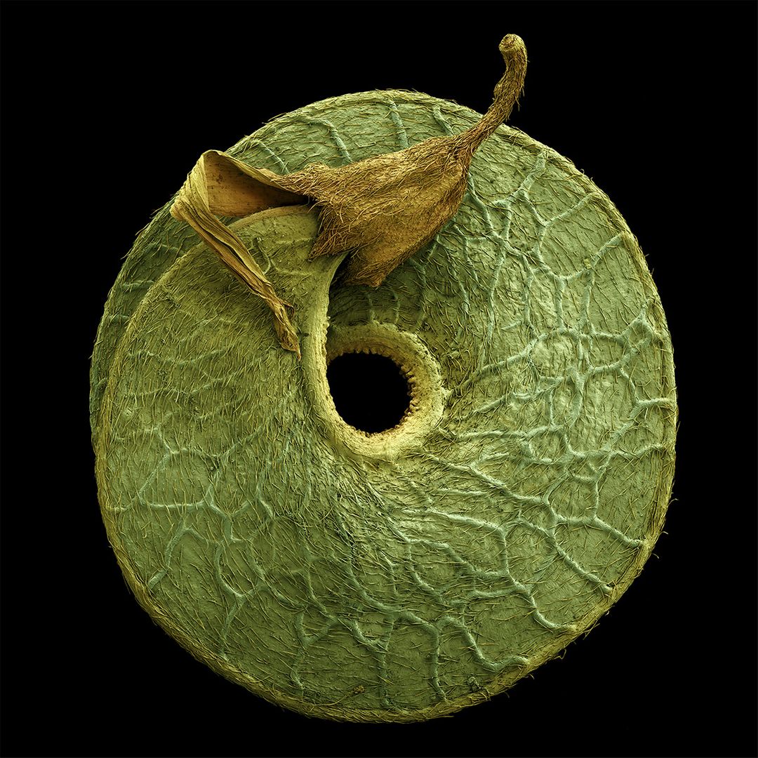
Colored #Micrographs Reveal the Incredible Intricacies of #Pollen, #Seeds, and #fruits @mymodernmet #agriculture share your #research at bit.ly/2XoPNAs #farmersconvention #Conference #agronomy #plants #technologynews #speakers #Spain #farming #Hydroponics #biotech
Something went wrong.
Something went wrong.
United States Trends
- 1. Bayern N/A
- 2. Mythos N/A
- 3. Madrid N/A
- 4. TACO N/A
- 5. Carreras N/A
- 6. Luis Diaz N/A
- 7. Olise N/A
- 8. Neuer N/A
- 9. Harry Kane N/A
- 10. 25th Amendment N/A
- 11. Vini N/A
- 12. Trent N/A
- 13. Vance N/A
- 14. Shane Smith N/A
- 15. #One_Two_CONNECT_Our_7 N/A
- 16. Martinelli N/A
- 17. #ChampionsLeague N/A
- 18. Upamecano N/A
- 19. Ghost Murmur N/A
- 20. DO SOMETHING N/A