#opticalmicroscopy результаты поиска

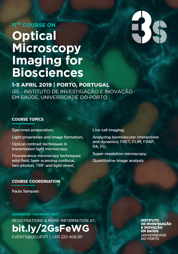
Searching for enlightenment in light microscopy? Apply to the Course on Optical Microscopy Imaging for Biosciences! Registration and more info: i3s.up.pt/training?v=116 #opticalmicroscopy #imaging #imageanalysis #fluorescence #science #i3s #ppbi


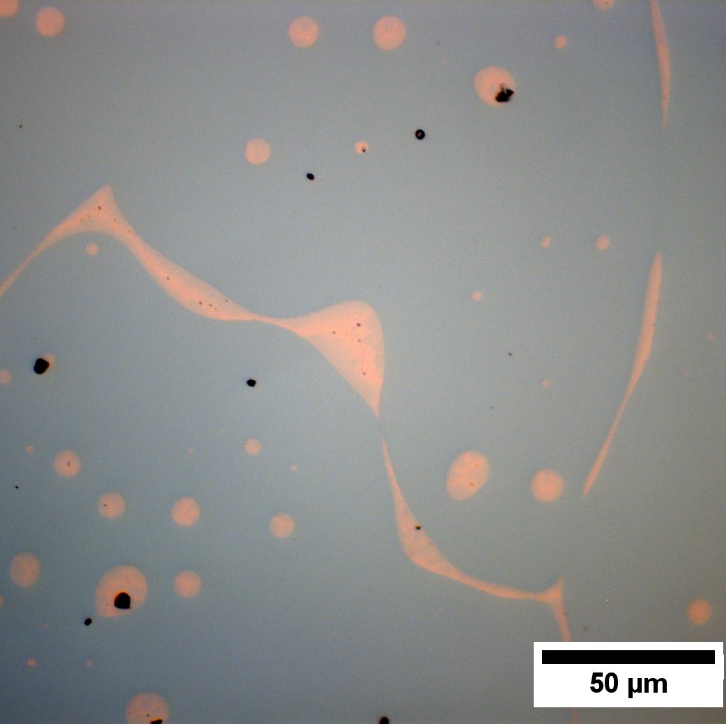
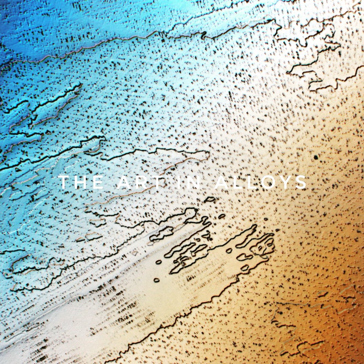
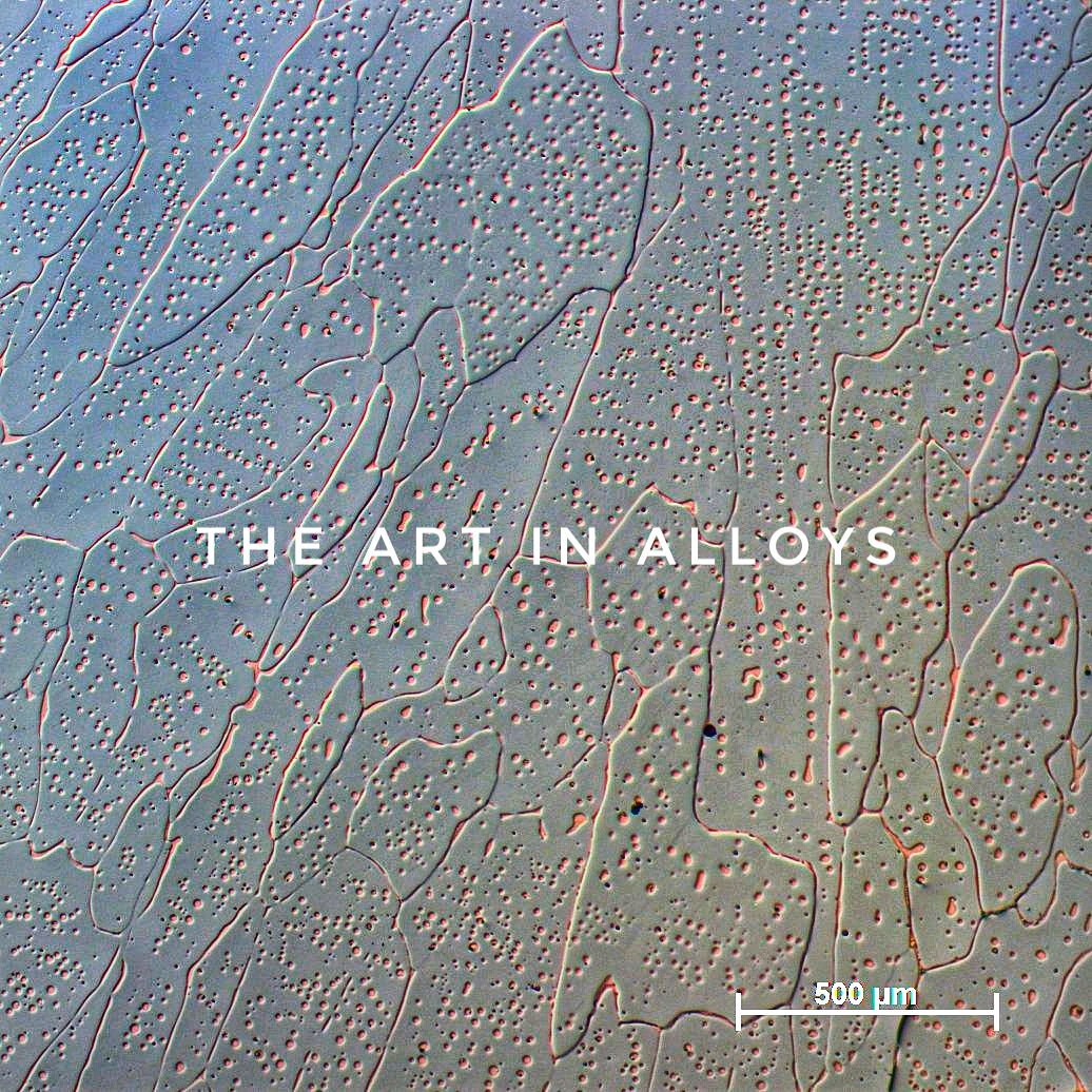
Friendly elements (in grey, all nicely mixed up) and a separatist a**hole. No recolouring. CoCrCuFeNi #highentropyalloy #opticalmicroscopy

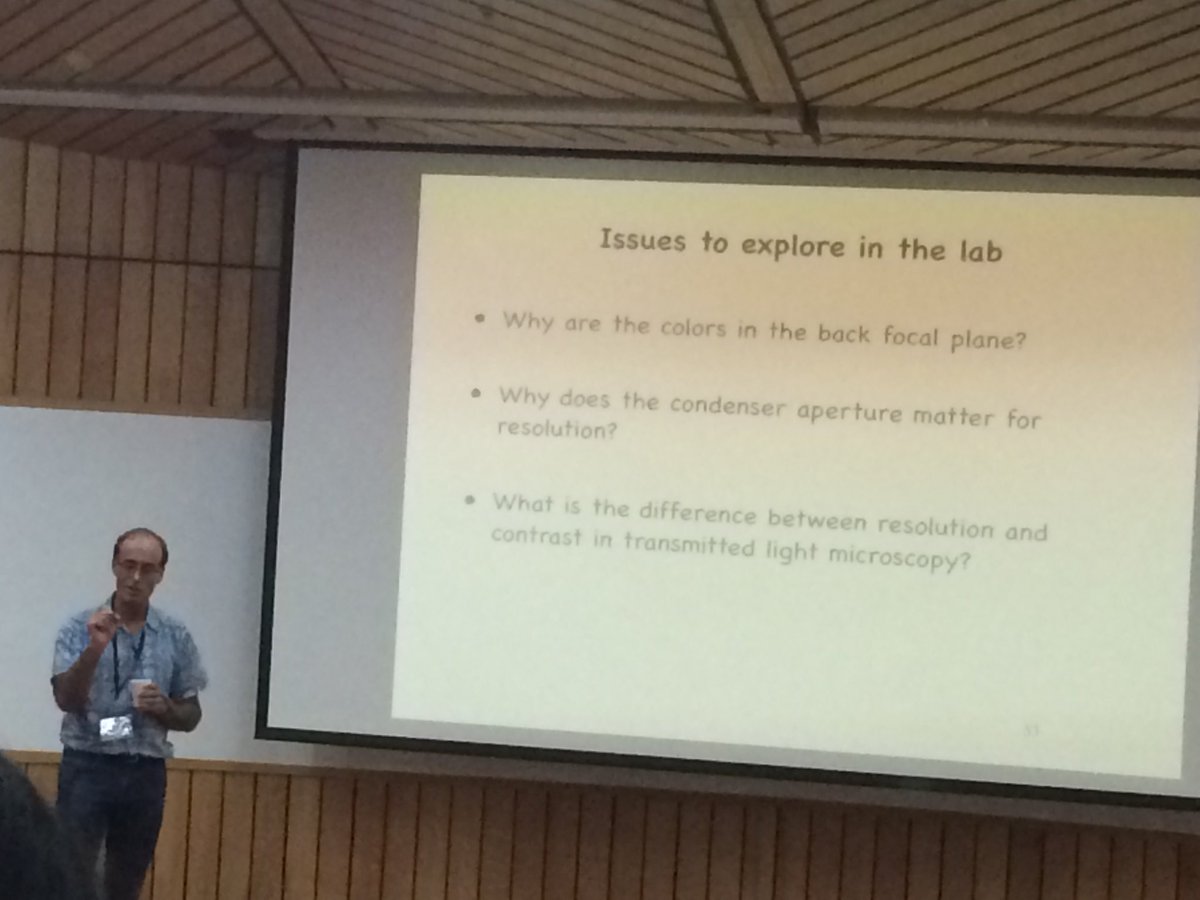
#ronvale explains the fundamentals of #opticalmicroscopy @NCBS_Bangalore #bmc2018 bangalore microscopy course

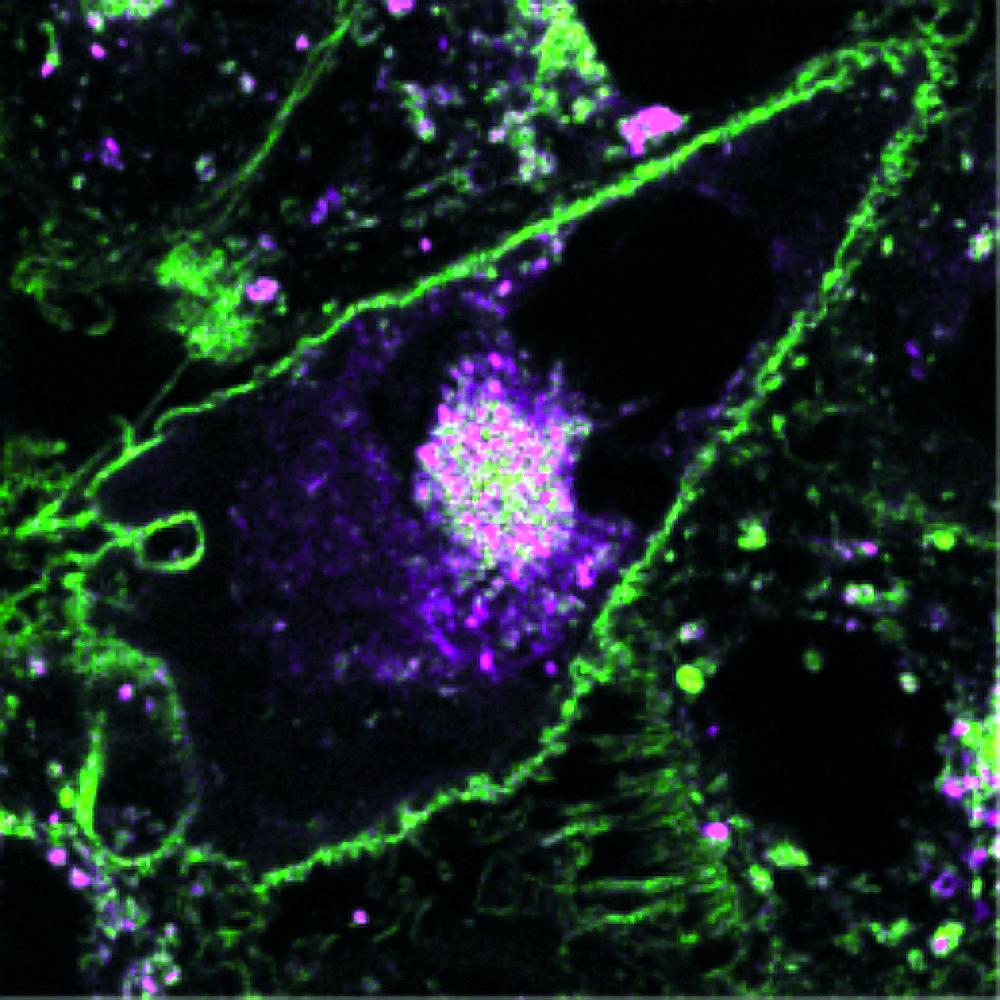
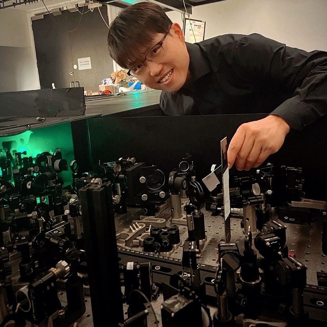
Hello there =). We are a #startup from @IITalk, pioneering new technologies in the field of #opticalmicroscopy. With @AD1959 and @VicidominiLab we are working on a new #superresolution #microscope based on #ImageScanningMicroscopy.
Book edited by Dr. @nirmal_mazumder Dept. of Biophysics and Dr. Gireesh Gangadharan, Dept. of Cell and Molecular Biology @MSLS_MAHE, @MAHE_Manipal titled “Advances in Brain Imaging Techniques” is published by Springer Singapore. Congratulations #BrainImaging #opticalmicroscopy

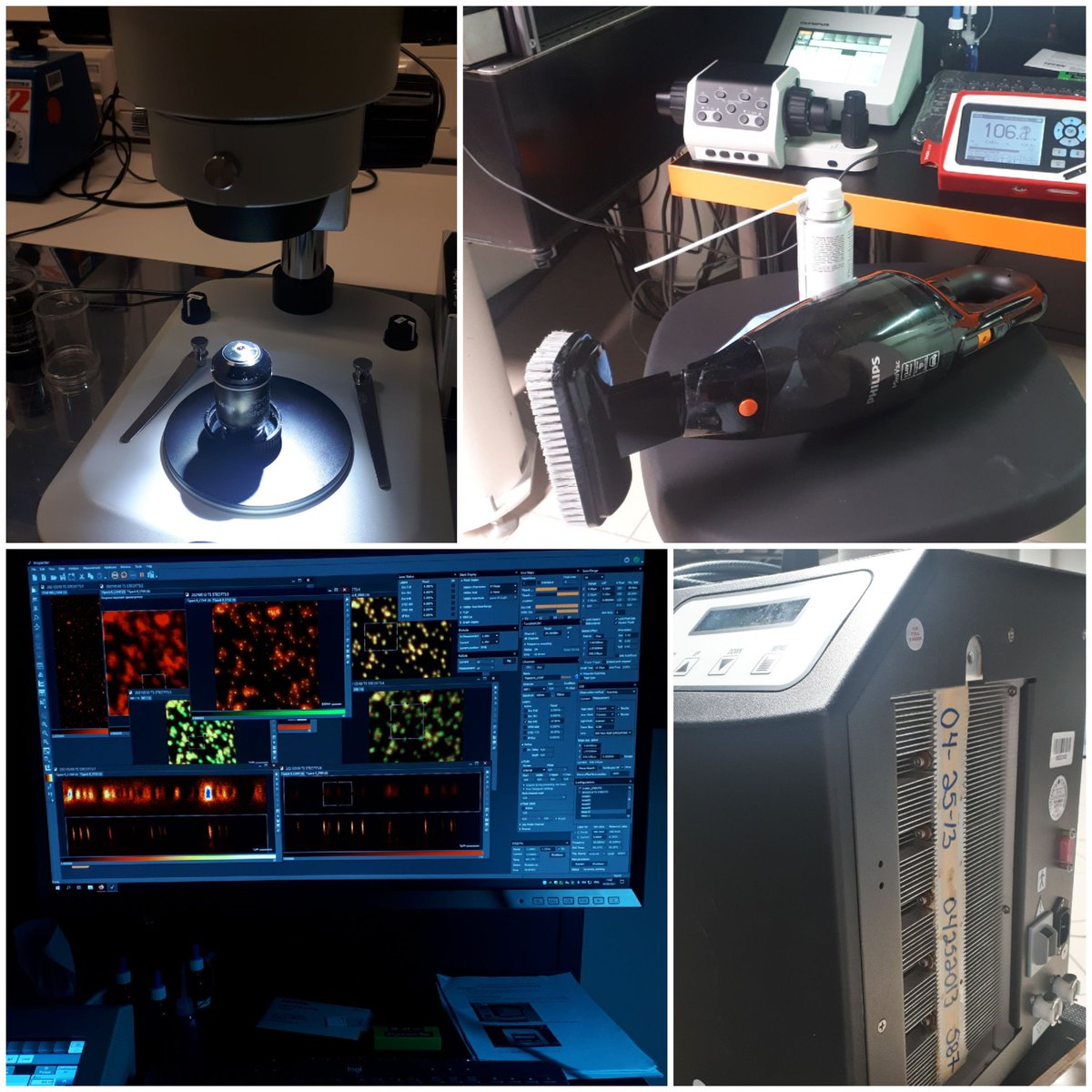
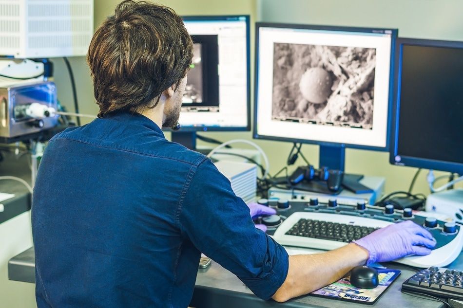
Time to thoroughly check the performances of our #nanoscopes, their 18 lasers, remove some dust and oil, and replace water in chillers! #facilitylife #opticalmicroscopy

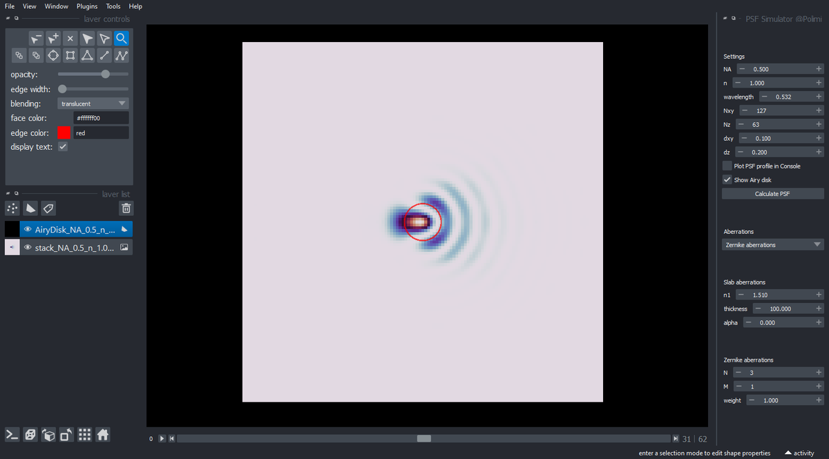
Managed to deploy my first #napari plugin! Just a 3D PSF generator, but a good exercise to see the potential of @napari_imaging for #opticalmicroscopy and #biophotonics. Many more to come. github.com/andreabassi78/…
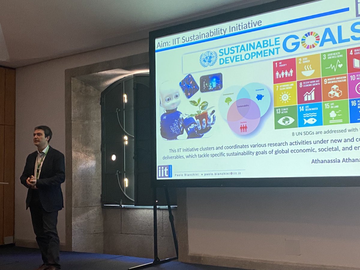
#touli @diasprolab @FOMconferences @IITalk #FOM2023 Paolo Bianchini on Sustainable Developments using #opticalmicroscopy #labelfree #water


Via #OPG_BOEx: UV curable resin as a rapid and superior sealant for STORM bit.ly/4qtHQqZ #OpticalMicroscopy #UVResin @SeoulNatlUni

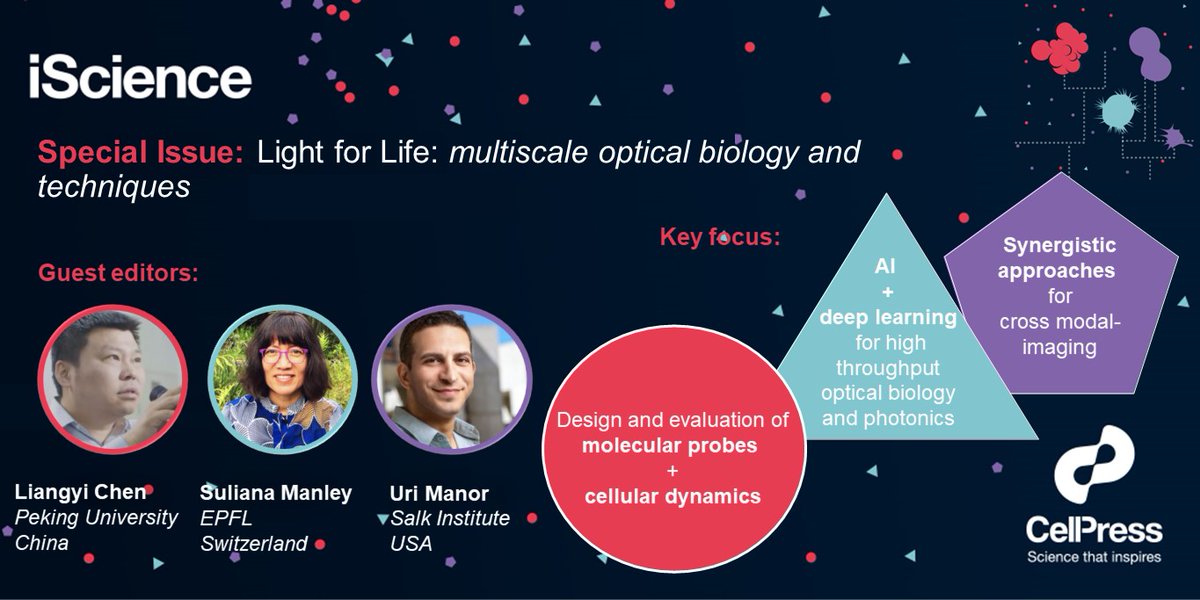
.@iScience_CP is interested in your research on emerging trends in #OpticalMicroscopy in the #SpecialIssue Light for life: multiscale optical biology and techniques. Guest edited by Uri Manor (@manorlaboratory), Suliana Manley (@SulianaManley) & Liangyi Chen (@Orangeroad2017)

#Nanotech #NanoOptics #OpticalMicroscopy: researchers discover that a nanomaterial -titanium oxynitride- allows a #superlens to reach an ultrahigh spatial resolution of 8 nm and 80 nm in the near-field and far-field, respectively - kpfu.ru/eng/news-eng/n…

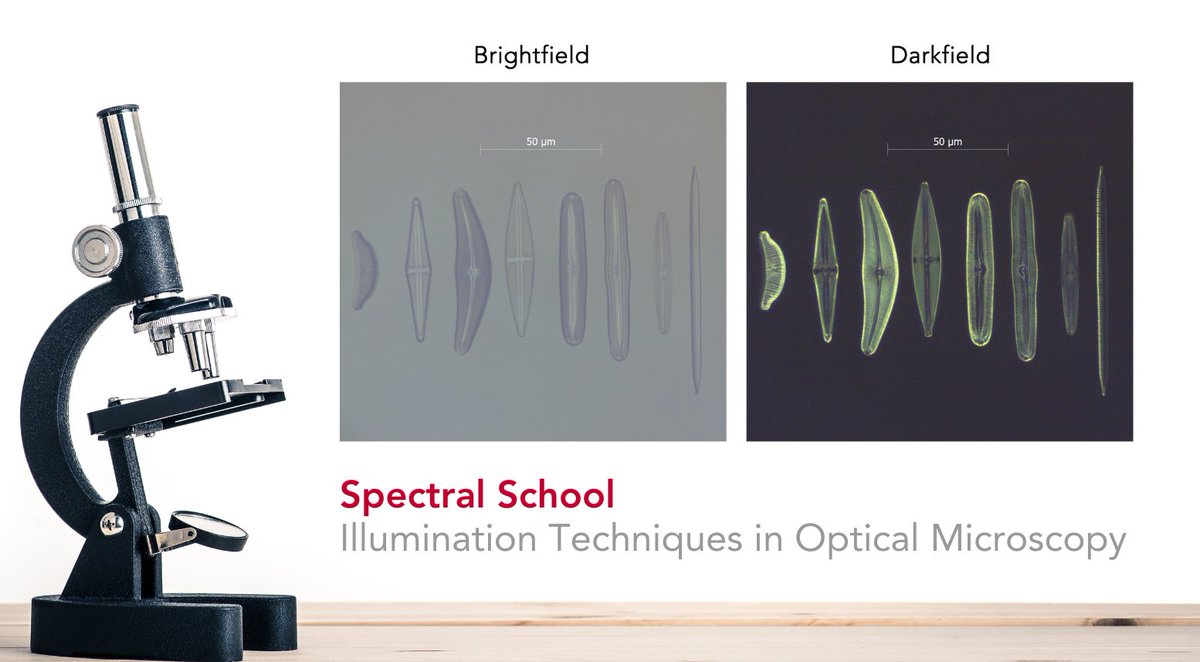
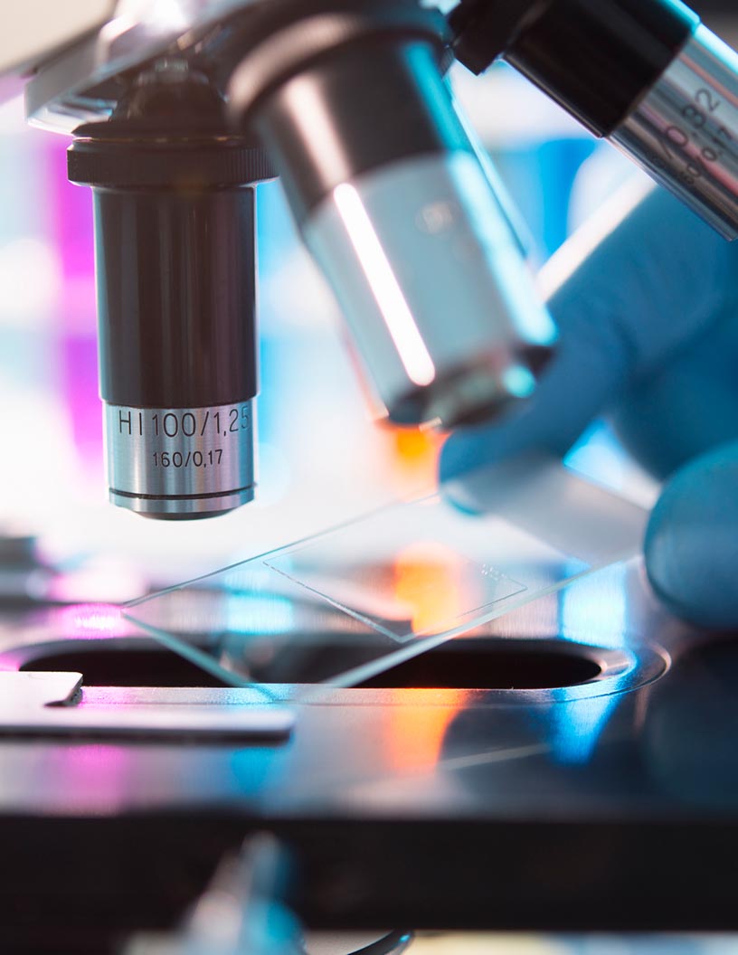
Ever wondered how scientists capture such detailed and high-contrast images of their samples under the microscope? 🤔 Read our latest blog to discover some of the most common illumination techniques in optical microscopy 🔬ow.ly/6ALo50P6658 #OpticalMicroscopy
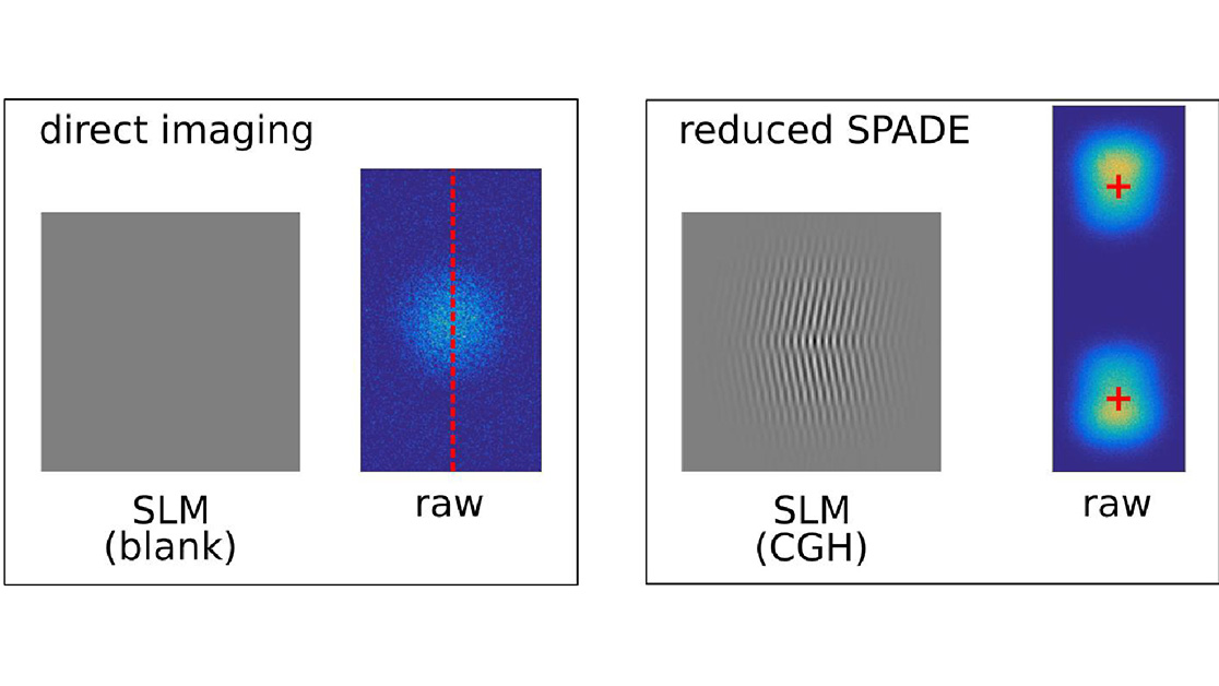
Via #OPG_OPTCON: Locating a single point source of light using an analytic estimator enabled by spatial mode demultiplexing bit.ly/4olpK8S #OpticalMicroscopy #Demultiplexing

#NobelLaureate & #LiNo17 attendee Stefan Hell explained the "Resolution Revolution" of #opticalmicroscopy at #LiNo16 ow.ly/74GC30cCxwJ

Via #OPG_OPTCON: Locating a single point source of light using an analytic estimator enabled by spatial mode demultiplexing bit.ly/4olpK8S #OpticalMicroscopy #Demultiplexing
Via #OPG_BOEx: UV curable resin as a rapid and superior sealant for STORM bit.ly/4qtHQqZ #OpticalMicroscopy #UVResin @SeoulNatlUni

#upbcampus Check out our – the Photon-X Spectrum Lab, led by Dr. Ștefan G. Stanciu – latest research in the near-field #opticalmicroscopy : “Diffraction-induced Artifacts in Scattering-type Scanning Near-field Optical Microscopy due to Lateral and Longitudinal Inhomogeneities”.

📢 Read our newest publication "Development of Projection Optical Microscopy and Direct Observation of Various Nanoparticles" by Toshihiko Ogura 🔗 Read the full article here: lnkd.in/eNDKns_y #Optics #Nanomaterials #OpticalMicroscopy #NewArticle
📸Highlights from #FOM2025 in #Taipei! We're having a great time connecting with researchers & showcasing ibidi's latest in #opticalmicroscopy🔬 🧫Thanks for stopping by Booth #4! Still time to visit & explore our innovative #solutions. #microscopy #research #innovation
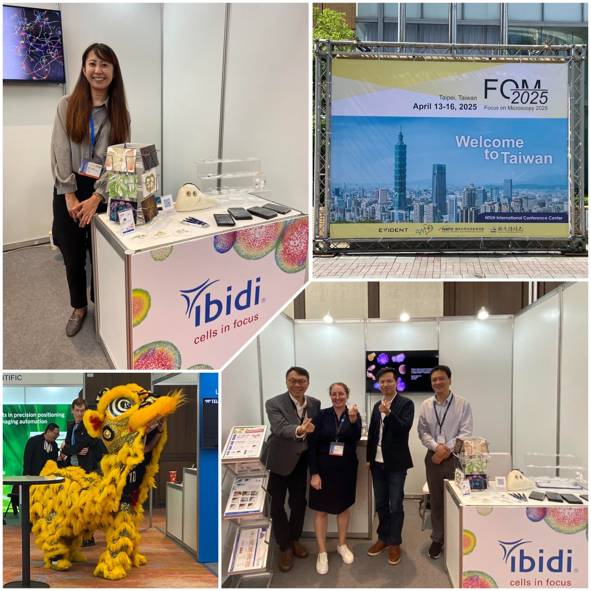
🎪 We’re heading to #FOM2025 in Taipei from April 13–16! Meet us at Booth #4 to explore ibidi’s latest in #opticalmicroscopy🔬for #biology & medicine. 🥼Dr. Irina Hein will be there to chat #research & #innovation. 🇹🇼See you in Taipei! #microscopy #ibidi #scienceinnovation


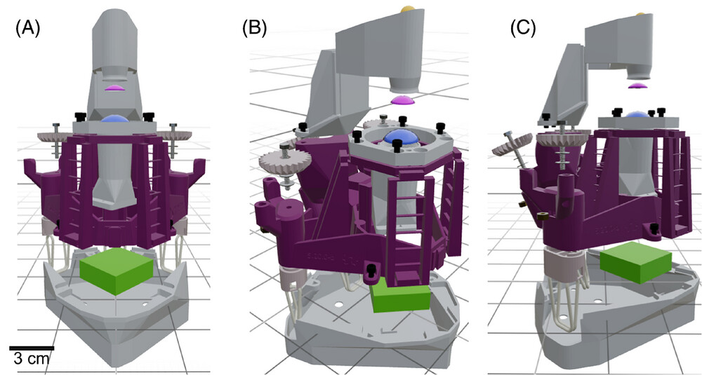
3D-printed microscope revolutionizes imaging! Affordable, customizable, and perfect for education, research, and diagnostics in low-resource areas. 🌍🔬 ➡️ow.ly/TMGw50VpvpN #OpticalMicroscopy #HistologicalImaging Image ©Christopher, J. et al. / J. Microsc.
Our comprehensive #microscopy equipment and #reagents eBook is your essential resource to make informed decisions when selecting the best microscopy tools for your lab. 🔗Download your free copy here: eu1.hubs.ly/H0hknDv0 #OpticalMicroscopy #ElectronMicroscopy #ScienceGuides


Looking for post. key words: optical measurement, optical microscopy, optical vortex, single-pixel imaging #opticalmeasurement #opticalmicroscopy #opticalvortex #singlepixelimaging
Looking for collaborators in the field of optical measurement (optical microscopy, laser engineering). #collaborator #opticalmeasurement #opticalmicroscopy #researcher

Scientists in the Rao group have developed a safer method to accelerate Phase Transitions in semiconducting 2D Materials, detailed in their paper published in Nature. Read the paper here: buff.ly/4dURvA6 #optoelectronics #OpticalMicroscopy #Crystallography
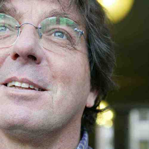
Cheers to the 2024 Enrico Fermi Prize winners for #opticalmicroscopy in biology! Prof. Alberto Diaspro @AD1959 and Prof. Francesco Saverio Pavone @fs_pavone! This recognition is well deserved for their outstanding contributions. fcld.ly/fermiprize

#3D #OpticalMicroscopy Market Set for Growth Surge: Rising Demand and Technological Innovations Drive Expansion 📈🔬. Explore the segments and factors propelling this market forward: rb.gy/lk31c8 #TechInnovation #SurfaceMeasurement #AutomotiveTech

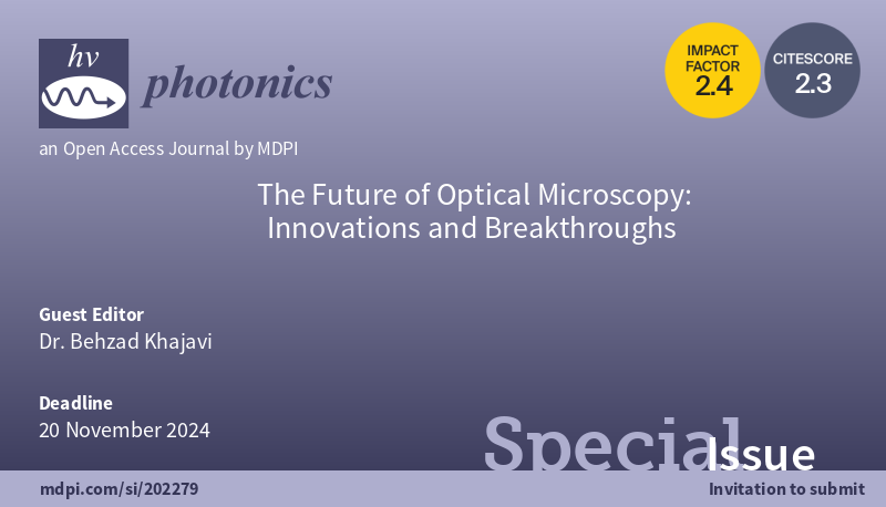
✨New Special Issue: "The Future of #OpticalMicroscopy: Innovations and Breakthroughs" 🔗mdpi.com/si/202279 📚 Edited by Dr. @khajavibeh 📢 Submission deadline: 20 November 2024 #Optics #Photonics
Searching for enlightenment in light microscopy? Apply to the Course on Optical Microscopy Imaging for Biosciences! Registration and more info: i3s.up.pt/training?v=116 #opticalmicroscopy #imaging #imageanalysis #fluorescence #science #i3s #ppbi
Via #OPG_BOEx: UV curable resin as a rapid and superior sealant for STORM bit.ly/4qtHQqZ #OpticalMicroscopy #UVResin @SeoulNatlUni
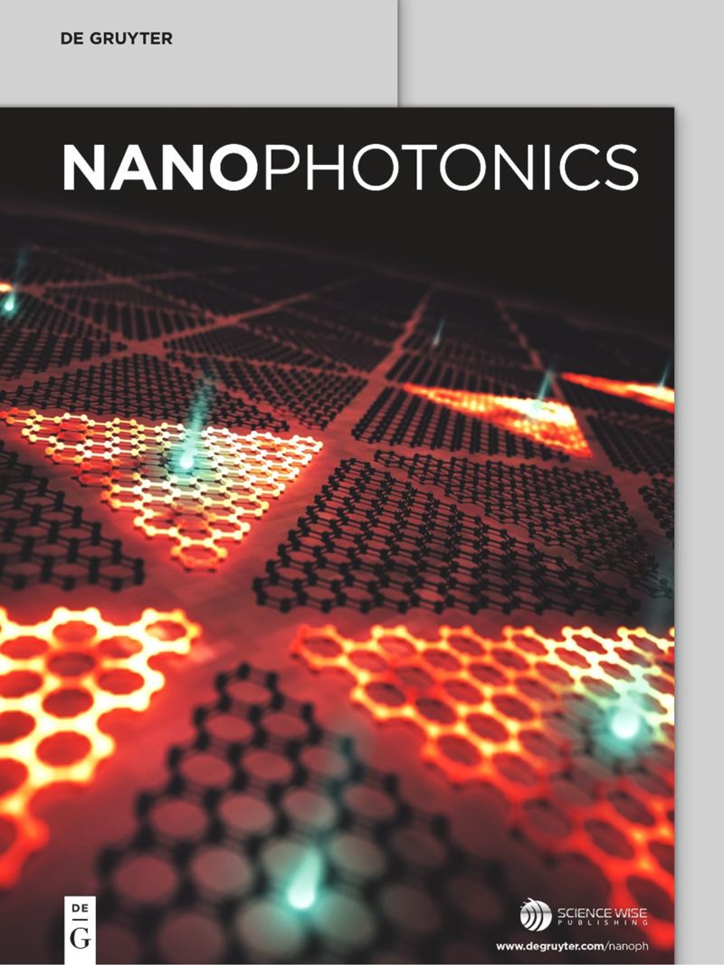
Sign up for ToC alerts for the latest on Near-field #OpticalMicroscopy and #NanophotonicDevices ... marketing.degruyter.com/nanophotonics?…
📢 Read our newest publication "Development of Projection Optical Microscopy and Direct Observation of Various Nanoparticles" by Toshihiko Ogura 🔗 Read the full article here: lnkd.in/eNDKns_y #Optics #Nanomaterials #OpticalMicroscopy #NewArticle
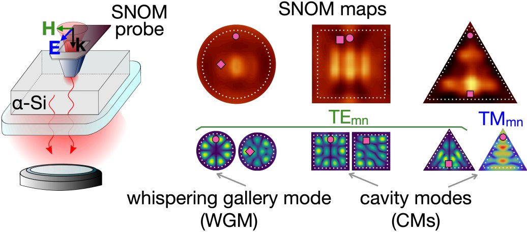
How all sets of subwavelength localized optical modes can be visualized in all-dielectric nanoantennas with a disk, square, and triangle shape? -> degruyter.com/document/doi/1… #nanophotonics #nanoantenna #opticalmicroscopy

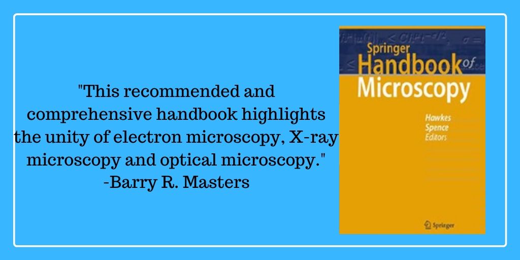
Check out OPN's latest book review: Springer Handbook of Microscopy @springerpub Read it here: ow.ly/IWl950zadU9 #electronmicroscopy #Xraymicroscopy #opticalmicroscopy #diffraction #optics #microscopy
#touli @diasprolab @FOMconferences @IITalk #FOM2023 Paolo Bianchini on Sustainable Developments using #opticalmicroscopy #labelfree #water
Our comprehensive #microscopy equipment and #reagents eBook is your essential resource to make informed decisions when selecting the best microscopy tools for your lab. 🔗Download your free copy here: eu1.hubs.ly/H0hknDv0 #OpticalMicroscopy #ElectronMicroscopy #ScienceGuides
Hello there =). We are a #startup from @IITalk, pioneering new technologies in the field of #opticalmicroscopy. With @AD1959 and @VicidominiLab we are working on a new #superresolution #microscope based on #ImageScanningMicroscopy.
Managed to deploy my first #napari plugin! Just a 3D PSF generator, but a good exercise to see the potential of @napari_imaging for #opticalmicroscopy and #biophotonics. Many more to come. github.com/andreabassi78/…
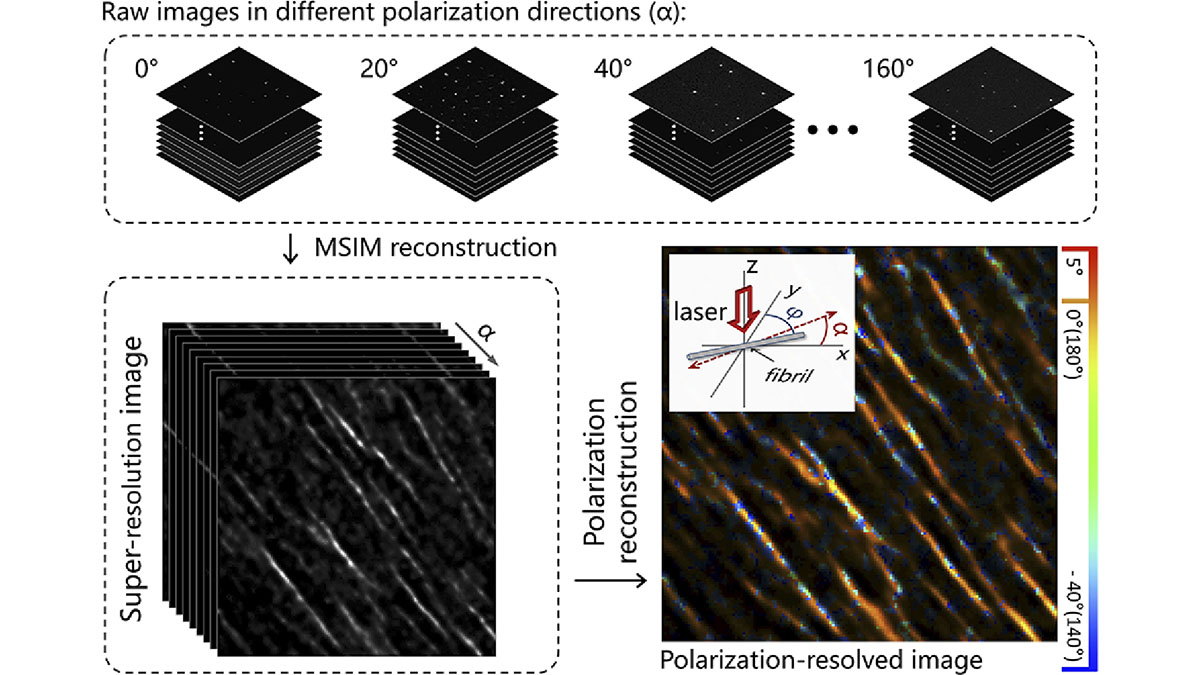
An Editors' Pick via #OPG_OL: Polarization-resolved super-resolution second-harmonic generation imaging based on multifocal structured illumination microscopy ow.ly/7Bam50QXOhV #OpticalMicroscopy #CollagenResearch
Book edited by Dr. @nirmal_mazumder Dept. of Biophysics and Dr. Gireesh Gangadharan, Dept. of Cell and Molecular Biology @MSLS_MAHE, @MAHE_Manipal titled “Advances in Brain Imaging Techniques” is published by Springer Singapore. Congratulations #BrainImaging #opticalmicroscopy
.@iScience_CP is interested in your research on emerging trends in #OpticalMicroscopy in the #SpecialIssue Light for life: multiscale optical biology and techniques. Guest edited by Uri Manor (@manorlaboratory), Suliana Manley (@SulianaManley) & Liangyi Chen (@Orangeroad2017)
If you are thinking of attending the Course on "Optical Microscopy Imaging for Biosciences" @i3S_UPorto, don't miss out early bird registration until March 3rd! #ALM #OpticalMicroscopy #Imaging #ImagingforBiosciences @pm_sampaio More info at i3s.up.pt/content/traini…
Via #OPG_OPTCON: Locating a single point source of light using an analytic estimator enabled by spatial mode demultiplexing bit.ly/4olpK8S #OpticalMicroscopy #Demultiplexing

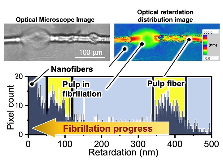
New optical system may enhance AI analysis of wood pulp quality insights.globalspec.com/article/15678/… #woodpulp #cellulosenanofiers #opticalmicroscopy
Something went wrong.
Something went wrong.
United States Trends
- 1. #SmackDown 24.4K posts
- 2. Mamdani 352K posts
- 3. Marjorie Taylor Greene 28.8K posts
- 4. Melo 14.8K posts
- 5. Aiyuk 4,329 posts
- 6. Kandi 6,818 posts
- 7. Azzi 5,328 posts
- 8. Myles Colvin N/A
- 9. Mama Joyce 2,423 posts
- 10. Sarah Strong 2,794 posts
- 11. #RissaHatchDay25 7,579 posts
- 12. Rebel Heart 1,504 posts
- 13. Hannah Hidalgo 1,623 posts
- 14. Congress in January 4,145 posts
- 15. joshua 56.9K posts
- 16. #OPNation N/A
- 17. Derik Queen 1,899 posts
- 18. #Dateline N/A
- 19. End 1Q N/A
- 20. Ilja 2,414 posts