JakobsLab
@JakobsLab
We are interested in #MitochondrialDynamics, #FluorescentProteins and #SuperResolutionMicroscopy! (Imprint: http://bit.ly/2tzaigd)
You might like


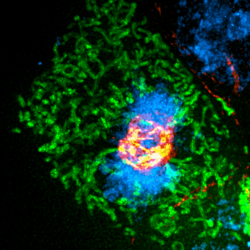
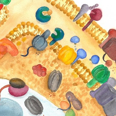
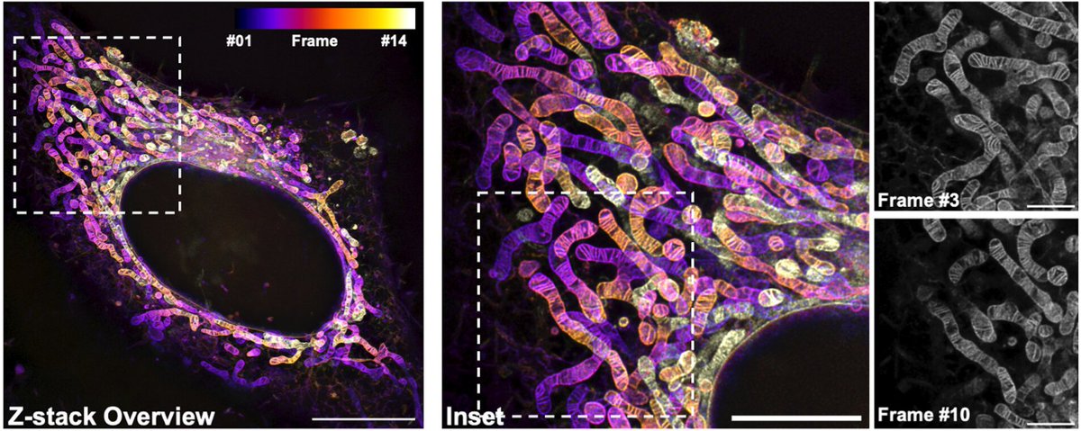
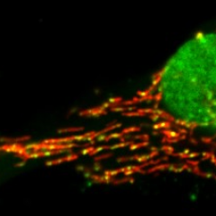
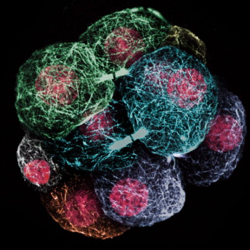
We got a belated Christmas present from @PNASNews: We made it onto the cover of the last issue for this year 😍 pnas.org/toc/pnas/curre… If you want to know more about the background, read our article about a new live-cell dye for mito imaging: doi.org/10.1073/pnas.2…


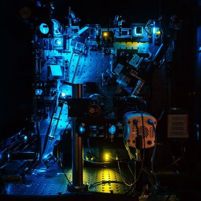
Thrilled to announce that our work on a STED simulation software designed for the validation of AI approaches and training of RL agents for autonomous control of STED imaging parameters is out now @NatMachIntell! @universitelaval @CERVO_ULaval @IID_ULaval nature.com/articles/s4225…
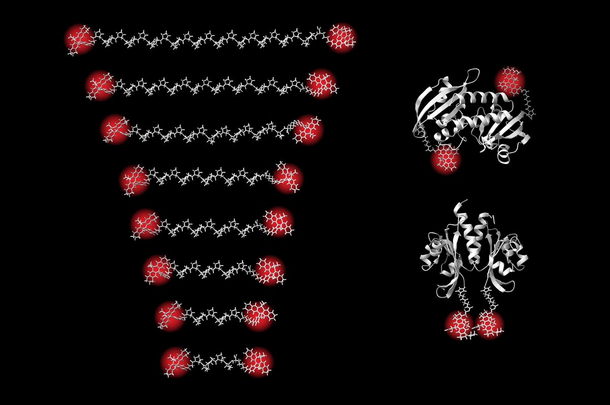
A team led by Steffen Sahl & @Stefan_W_Hell @mpi_nat and @mpi_mr_hd has succeeded in measuring distances within biomolecules using the #MINFLUX microscopy method, down to 1 nanometer and with Ångström precision. Read more in our press release: mpinat.mpg.de/4816514/pr_2419

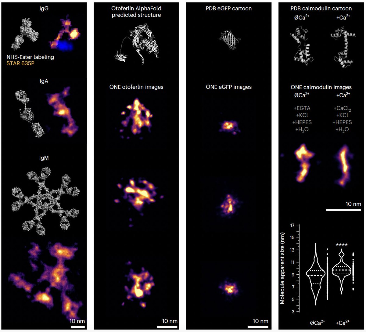
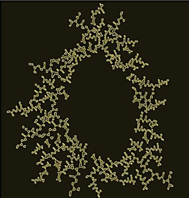
Protein shapes can now be studied using ONE🔬, a combination of ExM and #fluorescence fluctuation analysis. Happy to have contributed to this paper published in @NatureBiotech with many great scientists. Special thanks to @AliHShaib and Silvio Rizzoli. nature.com/articles/s4158…

Nanometer resolution in 3D with angstrom precision! Happy to have contributed to this study led by Steffen @StefanHellLabs @JakobsLab

Published in @ScienceMagazine: "Direct optical measurement of intramolecular distances with angstrom precision" (Sahl et al.) science.org/doi/10.1126/sc… @mpi_nat @mpi_mr_hd #Optics #Imaging #Microscopy #MINFLUX #Fluorescence
A nice talk from Stefan in PKU, with a lot of mitochondria, superresolution, and some twists to cryoEM in the end . Then we ended the day in the Emperior's diner for dinner😀 @JakobsLab @ZhixingChen2 @xipeng1


Beautiful work on the in situ structure of ATP Synthase by @dietrich_th from the Kühlbrandt group published today in @ScienceMagazine 🔬 Discover how this important protein complex shapes the inner mitochondrial membrane! 🧪 science.org/doi/10.1126/sc…
😍🤩
Super excited to share our latest work on the native in-cell organization of the mitochondrial respiratory chain 🥳 Using cryo-electron tomography🔬, we show how the respiratory complexes (and other complexes) are organized inside native mitochondria! biorxiv.org/content/10.110…

Want picture-perfect STED images of TOM & Co? -> follow our detailed protocol out today in Methods in Enzymology Vol. 710 "STED super-resolution microscopy of mitochondrial translocases" Great work together with @InamdarKaushik , @cdanieljans. @JakobsLab sciencedirect.com/science/articl…

Our #STED primer is out, you can learn about tips and tricks and current challenges in STED imaging

STED microscopy enhances the spatial resolution by reducing the effective fluorescence emission volume, disrupting the linear relation between excitation and spontaneous emission probability. Find out how in this weeks Primer: go.nature.com/4dD0PYX


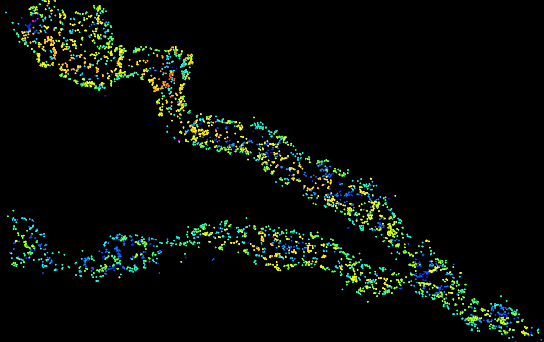
🚀Excited to share our latest paper, where we explore dynamic changes in mtDNA heteroplasmy in living cells! Discover how asymmetric partitioning and mitochondrial dynamics drive mtDNA variant segregation! 🔬embopress.org/doi/full/10.10… #Mitochondria #microscopy #microfluidics #mtDNA
🥳🚨Paper Alert🚨🥳 Excited to present our latest work on #Opa1 in this newly published paper @ScienceAdvances by @sahola6 and @LPazurek. We present here two new mouse models with impaired Opa1 processing. 1/🧵 science.org/doi/10.1126/sc… #mitochondria #mitochondrialdynamics @MPIAGE

I hope to see you all in Frankfurt at this exciting meeting! We promise, it will be worth the visit!

GBM Compact: Focus on Imaging Frankfurt, September 26-27, 2024 Deadline for early registration and abstract submission: June 30 gbm-compact.org 👇 👇 👇 👇 👇

GBM Compact: Focus on Imaging Frankfurt, September 26-27, 2024 Deadline for early registration and abstract submission: June 30 gbm-compact.org 👇 👇 👇 👇 👇
The GBM Compact conference „Focus on Imaging“ takes place from September 26-27 in Frankfurt! Early registration+abstract submisison is possible until June 30. Please visit the conference website for registration and further information: gbm-compact.org

"TFAM is an autophagy receptor that limits inflammation by binding to cytoplasmic mitochondrial DNA" nature.com/articles/s4155…
Are you interested in the intricate biology of fluorescent proteins? Do you want to push the power of MINFLUX microscopy even further? Live-cell imaging of mitochondrial protein dynamics tickles your brain? We want you 🧐 Check out the details below 👇 jobrxiv.org/job/the-max-pl…
Here comes our next gen PK Mito Orange Fix that can be fixed to IM for you to scrutinize the wonderland of mitochondria under STED, in a multiplexed way! Many thanks for the growing PK Mito team! @stephan_till @JakobsLab and Christian Juengst! pnas.org/doi/10.1073/pn…
After countless requests, we're thrilled to introduce PK Mito Orange FX - a fixable variant of PKMO. PKMO FX allows immunolabeling and CLEM approaches. Happy to have contributed to this project led by @ZhixingChen2 and Christian Jüngst. #mitochondria pnas.org/doi/10.1073/pn…

WHAT % OF YOUR TARGET BIOMOLECULES ARE YOU REALLY IMAGING? The *labeling efficiency* of your experiment is extremely important for image quality and quantitative results in super-resolution! Super excited about our new article in @naturemethods! 🤩 nature.com/articles/s4159…

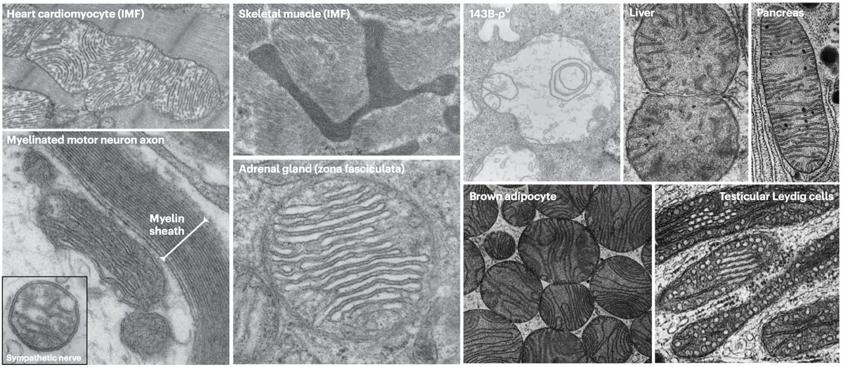
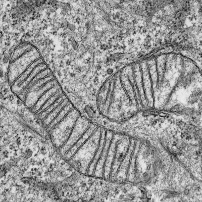
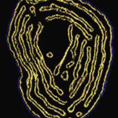
Mitochondrial research community - please post images of your favorite or most interesting mitochondrion Please indicate tissue, cell type, and other relevant information
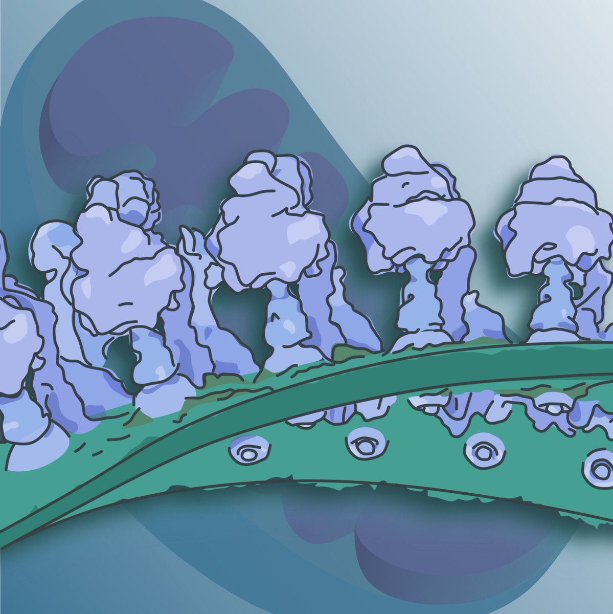
What do mitochondria look like? A little look at mitochondrial diversity by electron microscopy Source: picardlab.org/uploads/7/7/8/…


United States Trends
- 1. Massie 97.1K posts
- 2. #Varanasi 241K posts
- 3. Zvada N/A
- 4. #CollegeGameDay 2,254 posts
- 5. #MeAndTheeSeriesEP1 1.46M posts
- 6. Pat McAfee 1,250 posts
- 7. Todd Snider N/A
- 8. Lawson Luckie N/A
- 9. Aaron Donald 2,713 posts
- 10. #SaturdayVibes 5,614 posts
- 11. #Caturday 5,189 posts
- 12. Brooklynn 4,358 posts
- 13. Good Saturday 36.9K posts
- 14. Willie Green 5,179 posts
- 15. Charlie Becker N/A
- 16. James Franklin 2,300 posts
- 17. Marjorie 121K posts
- 18. Draymond 32K posts
- 19. Desmond Howard N/A
- 20. Wrigley Field 1,219 posts
You might like
-
 MITOtalks
@MitOtalks -
 Bewersdorf Lab
@bewersdorflab -
 Martin Picard
@MitoPsychoBio -
 PrudentLab
@LabPrudent -
 ReichertLab
@ReichertLab -
 Pagliarini Lab
@Pagliarini_Lab -
 Spirochrome
@spirochrome -
 Calì_Lab
@lab_cali -
 Payam A. Gammage
@mito_oncogene -
 Abberior
@Abberior -
 Alexis A. Jourdain
@jourdainlab -
 David Pla-Martín
@Dpla_ -
 Till Stephan
@stephan_till -
 Ruben QC
@RubenQC_Lab -
 Andreas Kohler
@AKohler_Mito
Something went wrong.
Something went wrong.