#gynpath search results

🔬 Pathology Basics 🧑🏫: Adenomyosis ~ Do you see the endometrial glands and stroma beneath the surface and deep in the myometrial wall? #GYNpath #Pathology #Histology #Uterus #PathologyBasics #PathResidents


🔬 Clear Cell Endometrial Adenocarcinoma ~ Images showing frequent clear cells with hobnailing (first image) and micropapillary growth pattern (second image) ~ #GYNpath #Pathology


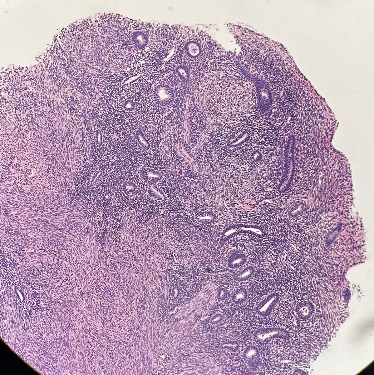
🔬📷 Fallopian Tube.. from a mile above! ✈️ (Super low power view) ~ #Pathology #Histology #GYNpath #PathArt

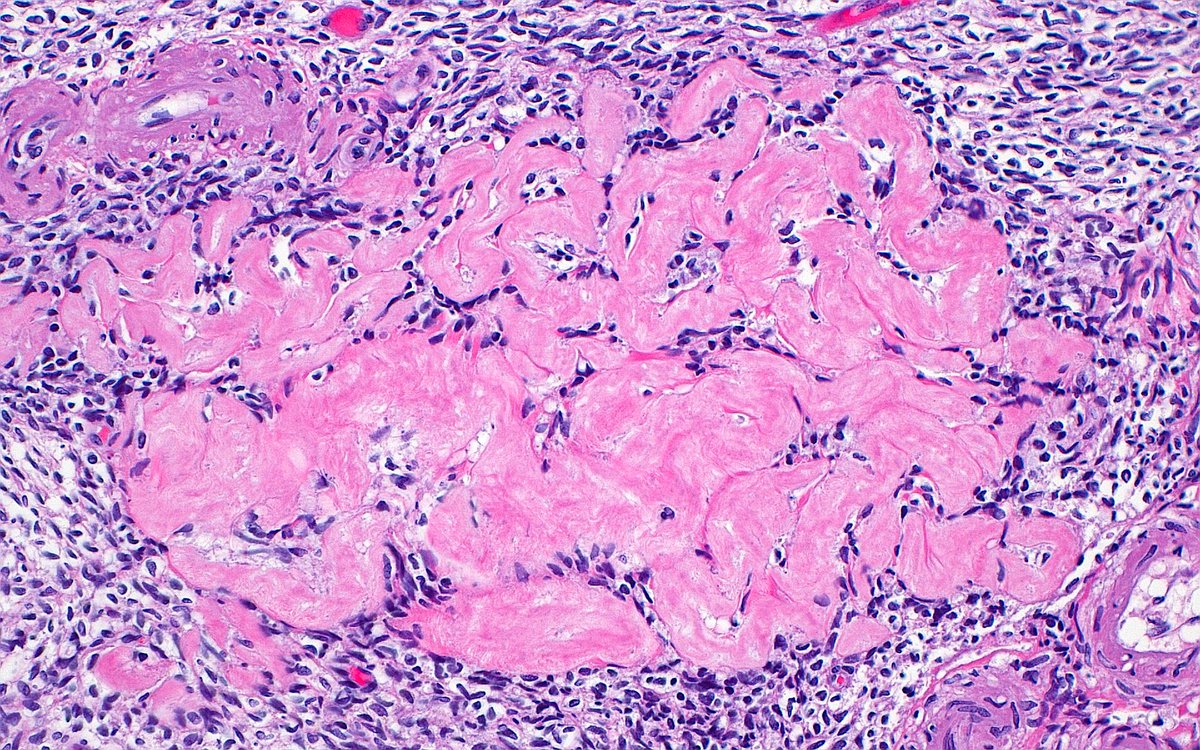
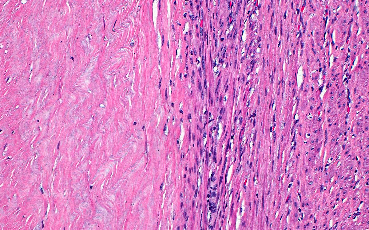
🔬 Leiomyoma with Hyaline Change ~ Collagen fibers (left half of image) next to smooth muscle cells (right half) that transitioned to an area of ischemic type necrosis (outside field of image) ~ #GYNpath #Pathology #Histology
🔬 Luteinized ovarian stromal cells (right) and spindled stromal cells (left) from a case of ovarian hyperthecosis ~ #GYNpath #histology #pathology


🚨Check out our new publication: The Spectrum of Vascular Lesions of the Upper Female Genital Tract: A Report of 55 Cases @md_kyle @MGBpathology @MGHPathology @IJGynP #GYNpath #BSTpath journals.lww.com/intjgynpatholo…


Incidental finding in the uterine corpus. What's the stain? What's the diagnosis? #gynpath #beautyinb9 #pathtwitter

🔬 Endometrial Endometrioid Adenocarcinoma ~ Back-to-back malignant glands (top two-thirds of image) above myometrium (bottom third of image) ~ #GYNpath #Pathology #Histology


Endometrioid carcinoma with secretory-like features may, at first glance, mimic benign secretory phase endometrium until you see the nuclear atypia and the architectural complexity next to the patient's age (older, postmenopausal female in this case) #PathTwitter #Gynpath


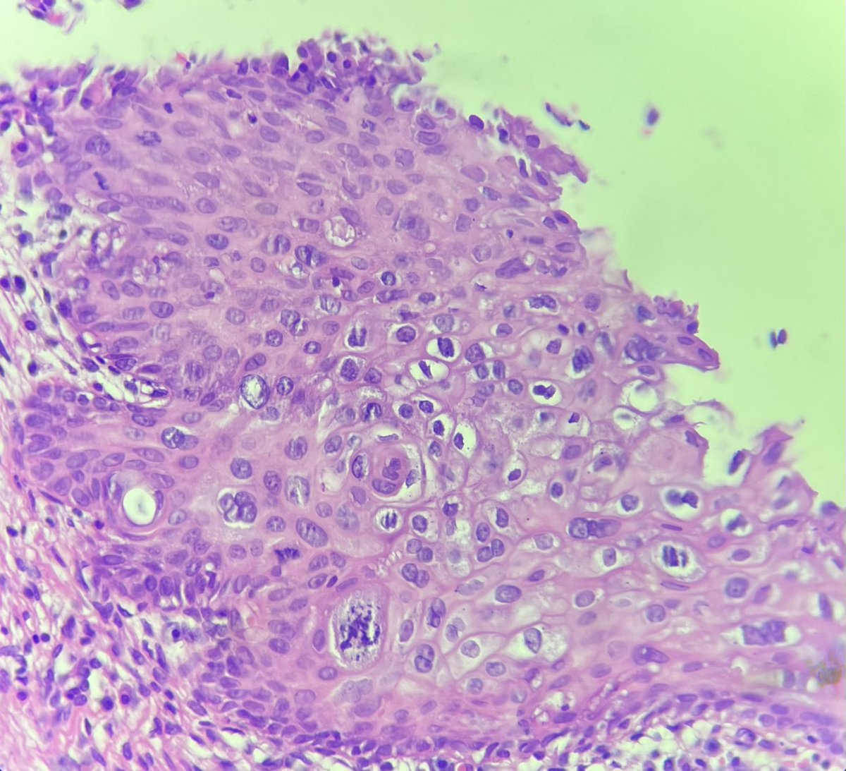
HPV-independent (differentiated) VIN. ➡️ Retained maturation ➡️ Atypia limited to basal and parabasal layers (🔺-shaped atypical basal keratinocytes with prominent intercellular bridges) ➡️ Hyperparakeratosis #IHCPath basally overexpressed, mutant p53 #PathTwitter #Gynpath

🔬 Molar Pregnancy ~ Trophoblastic cell proliferation around a villous from a complete hydatidiform mole ~ #GYNpath #PediPath #Pathology


Sometimes you can get an endometrial polyp Sometimes you can get an endocervical polyp Sometimes you can get an endometrial/endocervical polyp (arising from the lower uterine segment) Credit: Lisa Marinelli, MD #pathagonia #path4people #gynpath #pathx #obgyn #pathtwitter


🧬_______ missense mutation confirms the diagnosis. A) SMARCA4 B) DICER1 C) STK11 D) FOXL2 Hx: A 52-year-old woman with postmenopausal bleeding is found to have a large solid ovarian mass. #PathQuiz 🔬🧬 #Pathology #GYNPath #MolecularPath


📣 Check out the updated protocol: Reporting Results of Biomarker Testing of Specimens From Patients With Carcinoma of Gynecologic Origin 👉 doi.org/10.5858/arpa.2… #CAP #Archives #GYNPath

#breastpath #Gynpath #Molpath #Diagnexia Excelente oportunidad para ver a la Dra. Janira Navarro en un tema actual : Patología molecular práctica para neoplasias endometriales y mamarias 30 de abril de 2026 | 11:00 a. m. EST | 4:00 p. m. GMT | En español Una sesión enfocada en

Join Dr Janira Navarro for: Practical Molecular Pathology for Endometrial and Breast Malignancies April 30, 2026 | 11AM EST | 4PM GMT | En español A focused session on practical diagnostic considerations in Molecular Pathology for Endometrial and Breast Malignancies. ¡Webinar en

Endometriosis-associated neoplasms - review common diagnostic issues, and clinical, pathologic, and molecular characteristics here 👉 doi.org/10.5858/arpa.2… #CAP #Archives #GYNPath


New option for live-streaming our #gynpath #breastpath interactive digital slide-based course on May 23. Also FREE access for low-middle income country pathologists/trainees. Link to the digital slides will be sent for previewing in advance. Register: ucsf.cloud-cme.com/course/courseo…


New option for live-streaming our #gynpath #breastpath interactive digital slide-based course on May 23. Also FREE access for low-middle income country pathologists/trainees. Link to the digital slides will be sent for previewing in advance. Register: ucsf.cloud-cme.com/course/courseo…


Please join @ISGynP for our monthly gynecologic pathology journal club! April 15 at 12:00 US ET. This month's topic is Winning Abstracts from the USCAP Meeting. Register @ ISGyP.ca #PathTwitter #GynPath #GynaePath


🔬 Cracking the Code of Ovarian Mucinous Neoplasms - review diagnostic criteria, recent updates, and ongoing challenges here 👉 doi.org/10.5858/arpa.2… #CAP #Archives #GYNPath


Which specific marker is this? Diagnosis? #NeuroPath #GynPath #Pathology #PathTwitter #PathX #PathResidents #Pathologists #SurgPath

Announcing a free @ISGynP sponsored live virtual slide session on April 9, 2026 at 12 noon US ET by Dr. Salim on "Squamous Cell Carcinoma of the Cervix". Register now @ isgyp.ca #gynpath #gynaepath #PathTwitter
isgyp.ca
Home - ISGyP
Home - ISGyP
Excited that our paper on vascular lesions of the upper female genital tract was among the Top 10 most accessed articles in International Journal of Gynecological Pathology in 2025! 🎉 journals.lww.com/intjgynpatholo… @MGBpathology @IJGynP #SurgPath #GYNpath #BSTpath
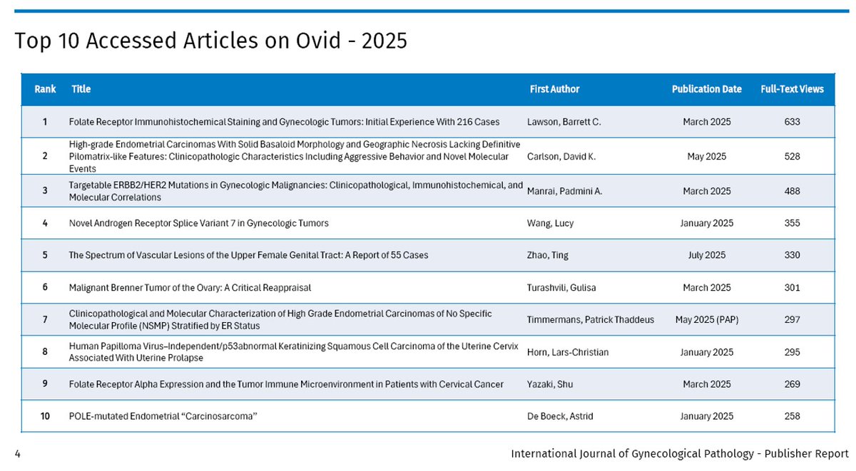
🚨Check out our new publication: The Spectrum of Vascular Lesions of the Upper Female Genital Tract: A Report of 55 Cases @md_kyle @MGBpathology @MGHPathology @IJGynP #GYNpath #BSTpath journals.lww.com/intjgynpatholo…
Metastases to the ovary can closely mimic primary ovarian tumors making accurate diagnosis both challenging and critical. Review key clinical, gross, and microscopic clues here 👉 doi.org/10.5858/arpa.2… #CAP #Archives #GYNPath


Please join us for our monthly webinar on Wed 8th April at 3 pm UK time. This month's topic is a session on 'The Clinically Important Diagnostic Thresholds in Gynae Pathology' with Dr Costigan. Free to register @ thebagp.org/events/list/ #PathTwitter #GynPath #Gynaepath


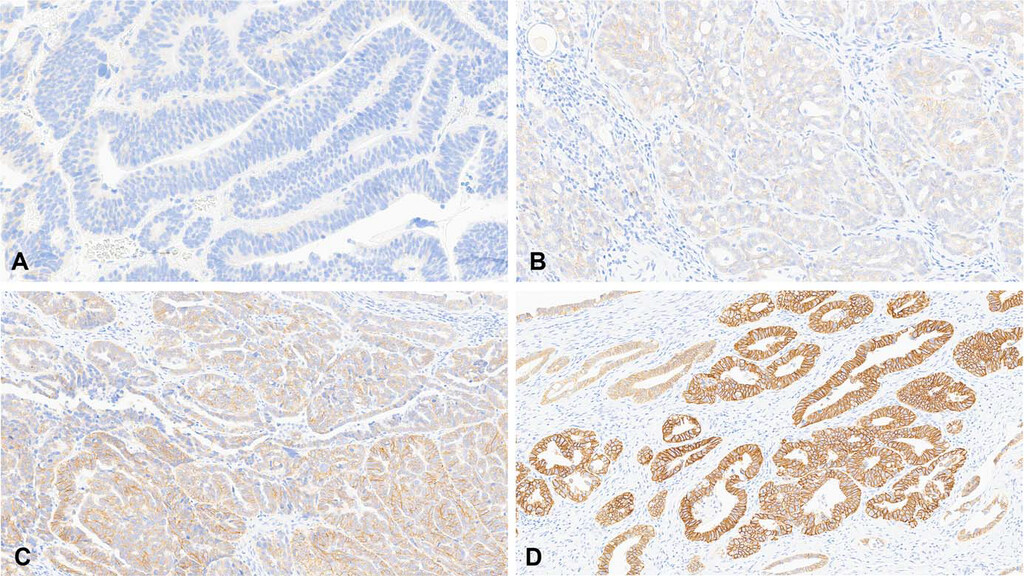
#Gynpath #Surgpath #pathresidents #Pathtwitter Some images of an endometrioid-type adenocarcinoma of the ovary in a 60-year-old woman. Mismatch repair proteins MLH1 and PMS2 show loss of staining (note negatively-staining tumor cell nuclei, with positively-stained stromal
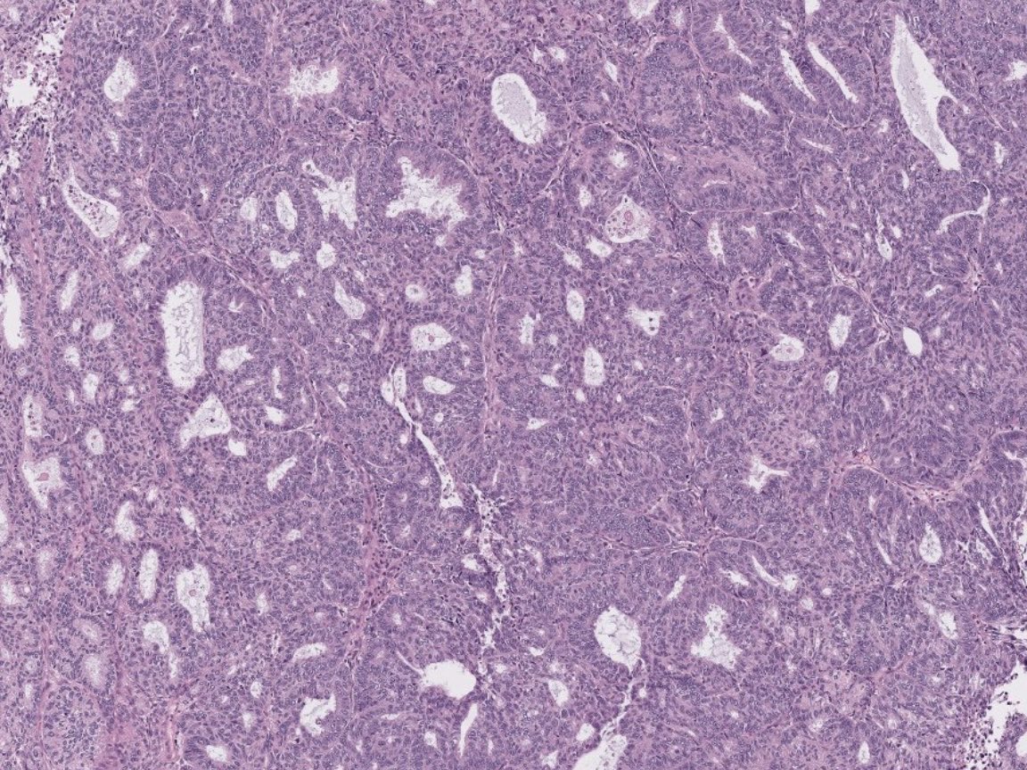
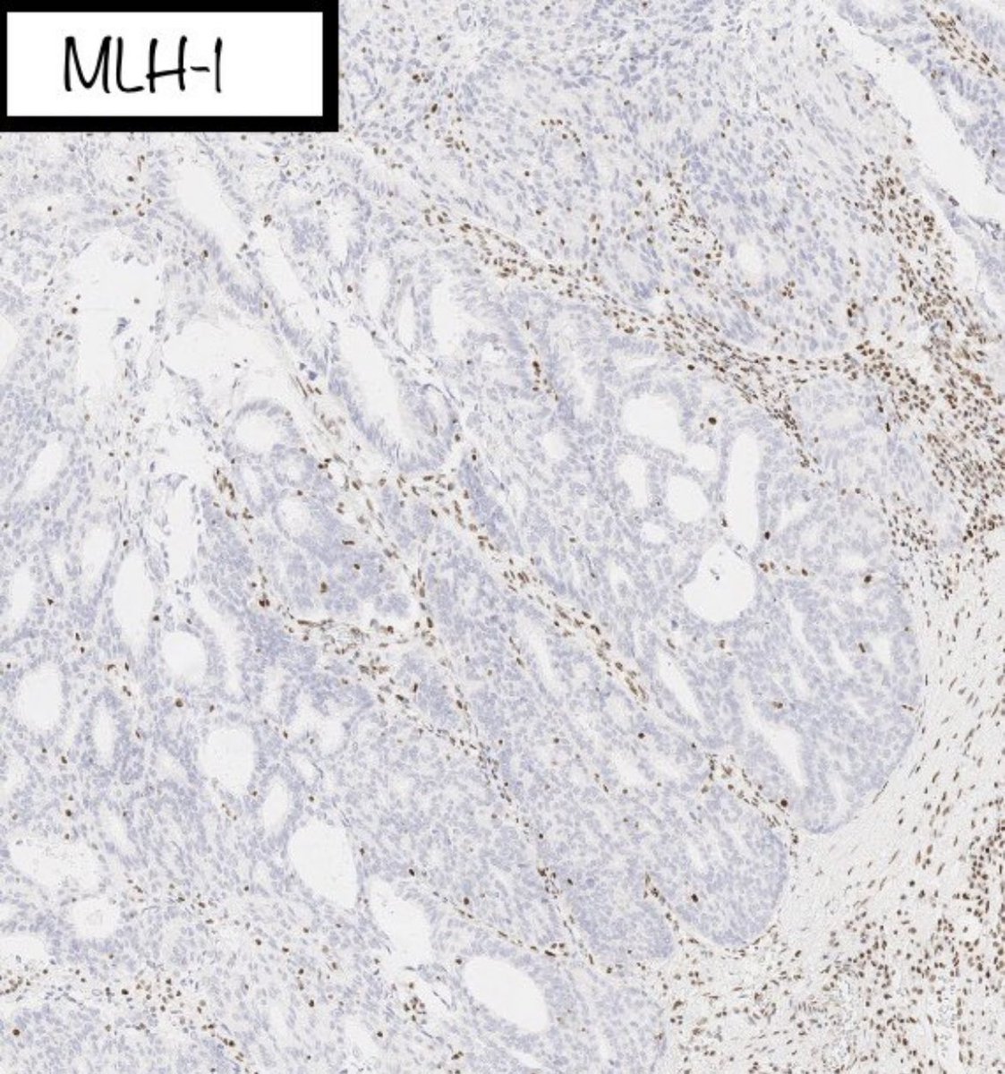
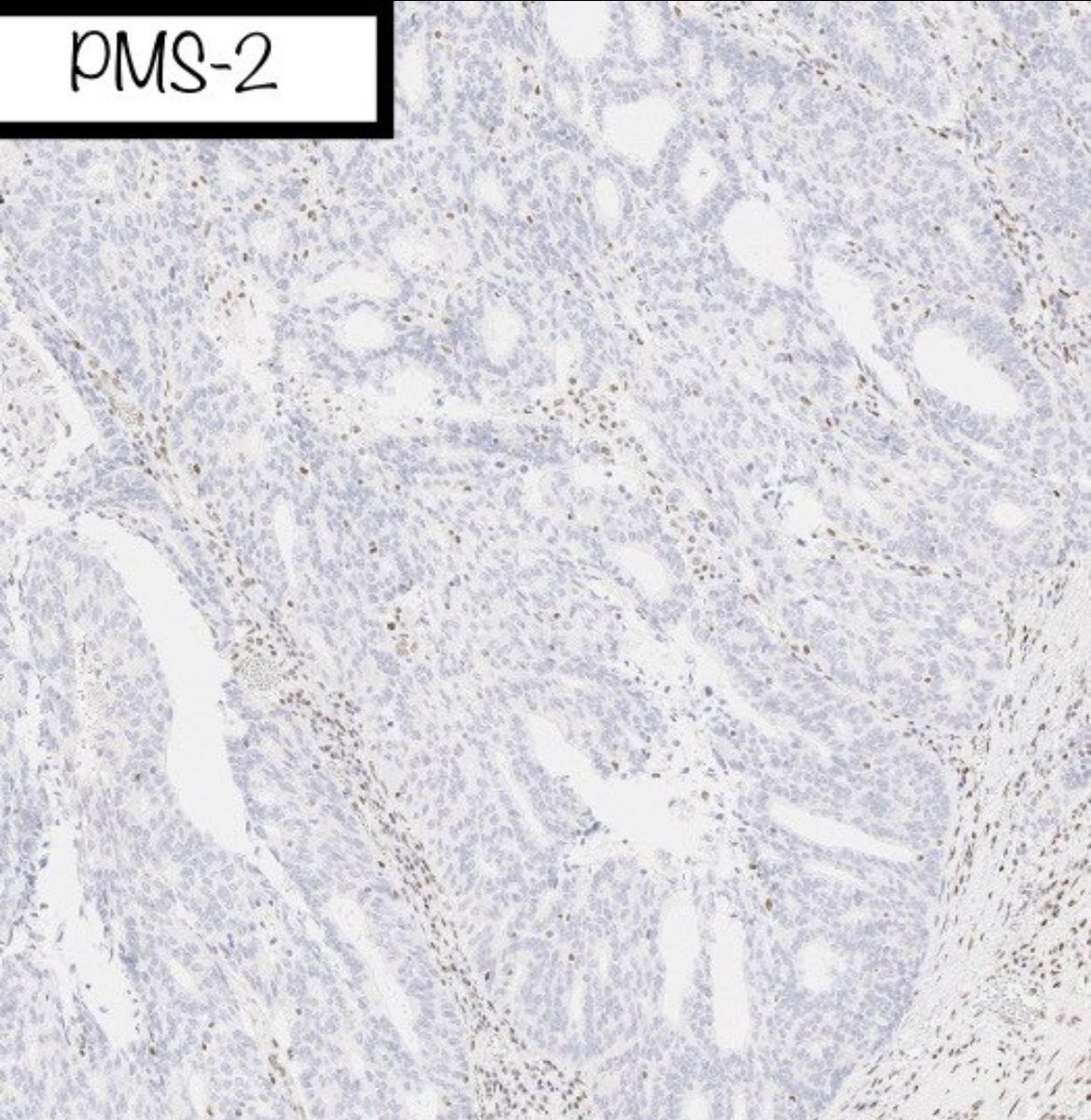
#Gynpath #Surgpath #pathresidents #Pathtwitter Some images of an endometrioid-type adenocarcinoma of the ovary in a 60-year-old woman. Mismatch repair proteins MLH1 and PMS2 show loss of staining (note negatively-staining tumor cell nuclei, with positively-stained stromal
Call For Applicants for Open Positions as ISGyP LiVE Moderators! Members of ISGyP are eligible to apply, each of which is a 2-year term. Closing date for applications April 24. For more details✉️[email protected] #Gynpath #GynaePath #PathTwitter


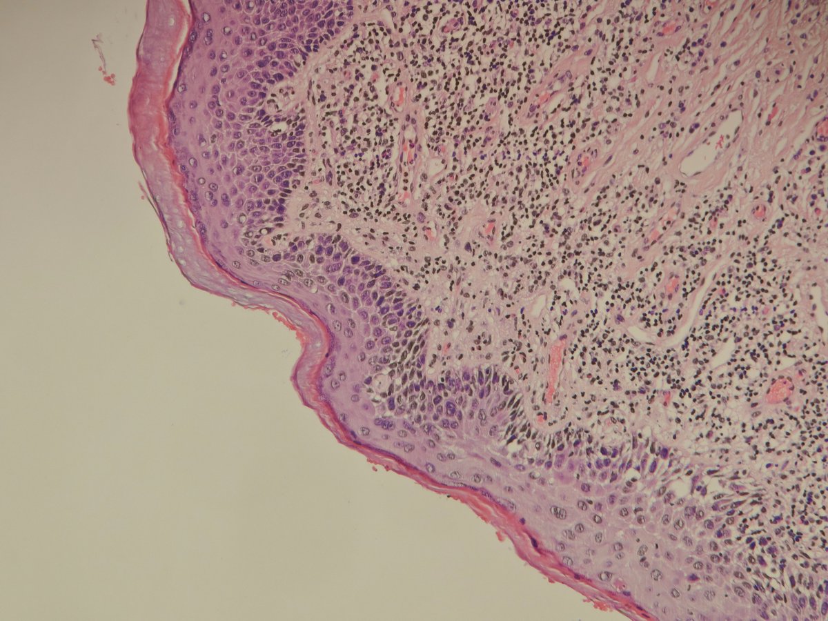
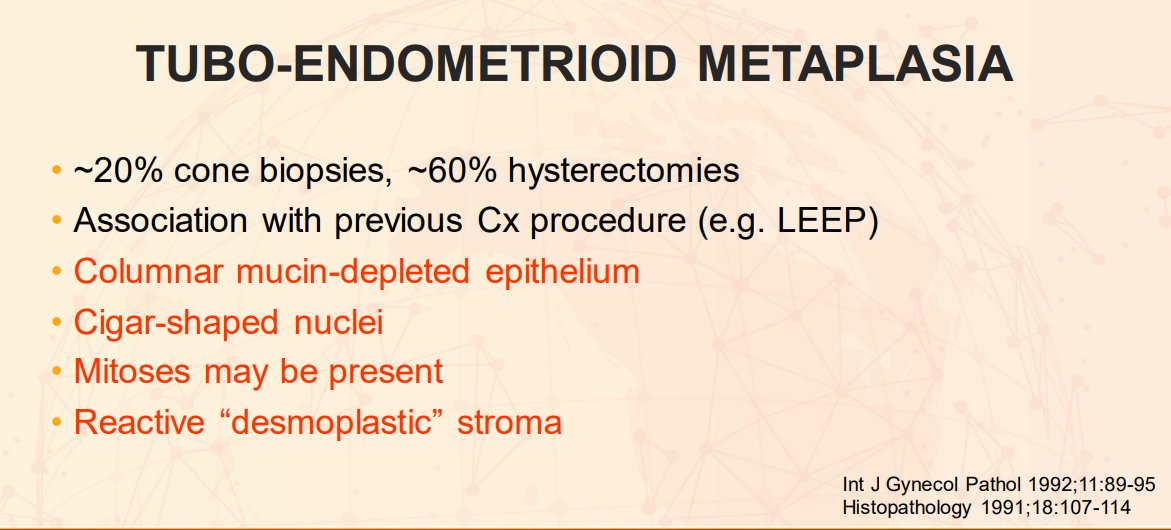
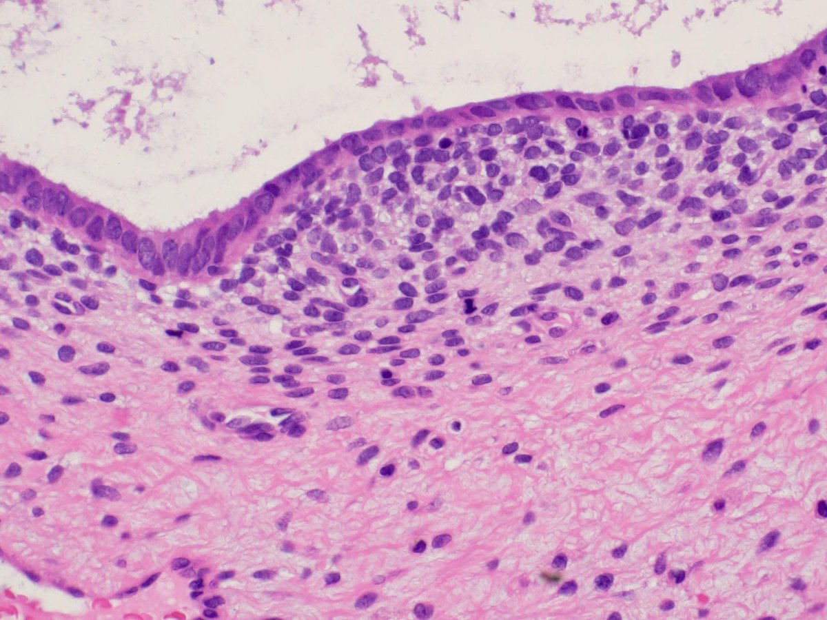
Uterine cervix - History of adenocarcinoma in situ (AIS) with positive margins → new biopsies come in Is this AIS? It’s tuboendometrioid metaplasia. ✅ Seen in ~20% of cone biopsies ✅ Often linked to prior cervical procedures ✅ Columnar mucin-depleted epithelium + reactive


A 62-year-old woman presents with a polypoid intrauterine mass. Aggressive cases of this tumor demonstrate: A) JAZF1::SUZ12 fusion B) FOXL2 mutation C) DICER1 mutation D) MYBL1 amplification #PathQuiz 🔬🧬 #Pathology #GYNPath #MolecularPath Clue in caption 🧐


An interesting incidental finding in a placenta! What do you think it is? Can you name the stain?😉 #PathTwitter #PathX #GYNPath #PediPath


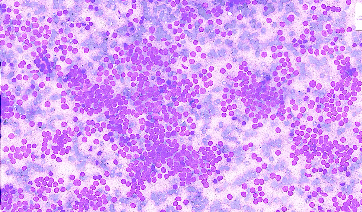
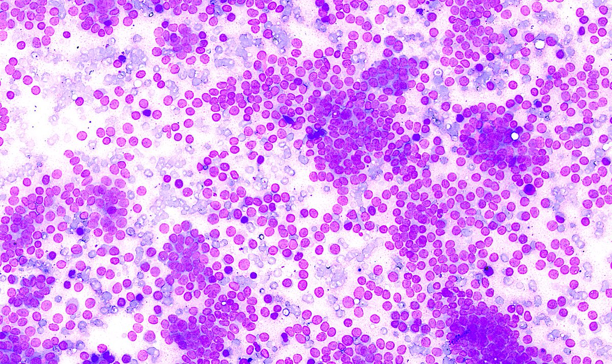
Adult female. Ovarian mass. Scrape smears prepared during the frozen section. #CytoPath #GynPath #CytoHisto correlation


I love all my children equally but WHOA do we have a treat for you! Please join us tomorrow morning @ 11 ET for our next ISIMM webinar: a #gynpath extravaganza. @AnneMillsMD: MMR/PD-L1 @BrookeHowittMD: mesenchymal tumors Jaclyn Watkins: vulvar pathology Matt Quick: endometrium


🔬 Luteinized ovarian stromal cells (right) and spindled stromal cells (left) from a case of ovarian hyperthecosis ~ #GYNpath #histology #pathology
🔬 Clear Cell Endometrial Adenocarcinoma ~ Images showing frequent clear cells with hobnailing (first image) and micropapillary growth pattern (second image) ~ #GYNpath #Pathology
HPV-independent (differentiated) VIN. ➡️ Retained maturation ➡️ Atypia limited to basal and parabasal layers (🔺-shaped atypical basal keratinocytes with prominent intercellular bridges) ➡️ Hyperparakeratosis #IHCPath basally overexpressed, mutant p53 #PathTwitter #Gynpath

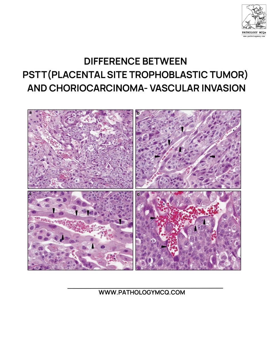
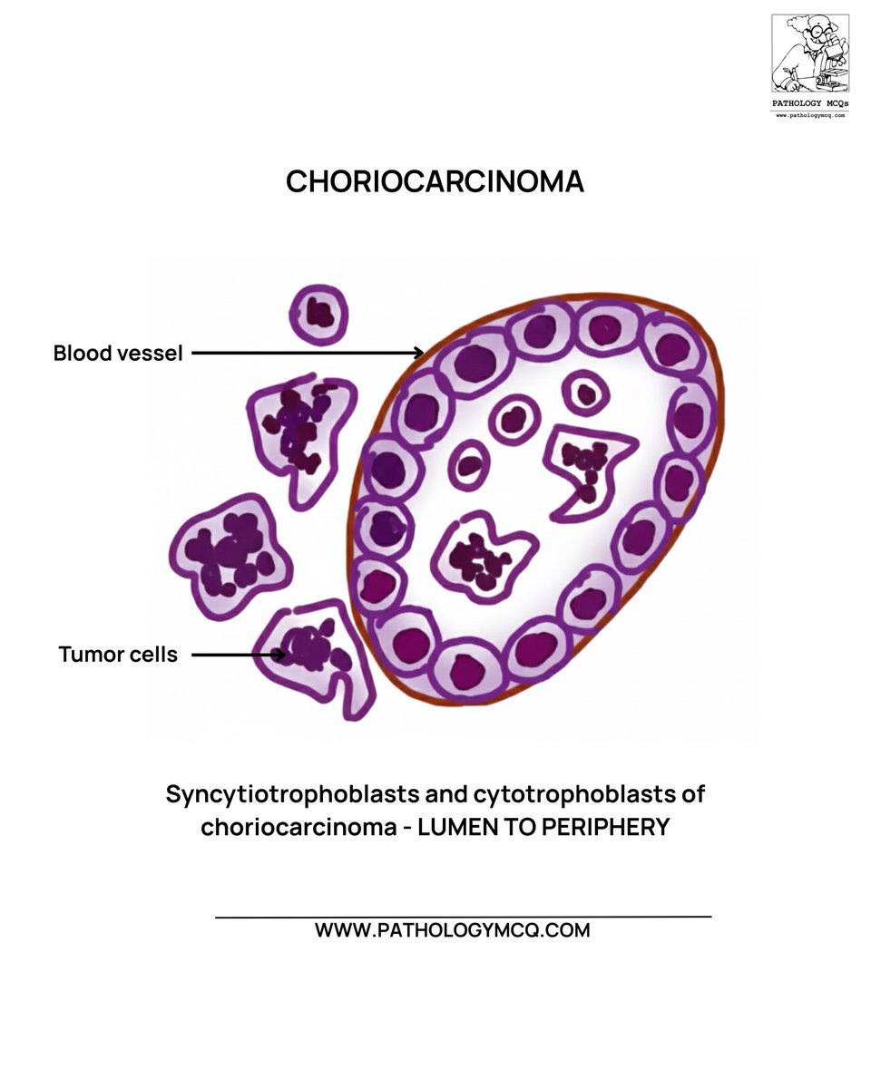
🔬 PSTT vs Choriocarcinoma — vascular invasion made simple! ➡️ PSTT: Periphery → Lumen ➡️ Choriocarcinoma: Lumen → Periphery Small clue, BIG diagnostic difference. 🩸✨ #Pathology #GynPath #Histopathology #MedEd #FRCPath #NEETSS


🔬📸 "Duos" 🥚🥚 ~ 2 Primordial Follicles (Top) - Normal Ovary ~ 2 Call-Exner Bodies (Bottom) - Granulosa Cell Tumor #Pathology #GYNpath #Histology #PathArt


Tender abdominal nodule. Cutaneous endometriosis. Dermis with scattered endometrial glands with stroma and hemorrhage and surrounding dermal fibrosis. The endometrium in skin also cycles throughout the month. #dermpath #gynpath #pathology


🔬📷 Fallopian Tube.. from a mile above! ✈️ (Super low power view) ~ #Pathology #Histology #GYNpath #PathArt
Incidental finding in the uterine corpus. What's the stain? What's the diagnosis? #gynpath #beautyinb9 #pathtwitter
🔬 Pathology Basics 🧑🏫: Adenomyosis ~ Do you see the endometrial glands and stroma beneath the surface and deep in the myometrial wall? #GYNpath #Pathology #Histology #Uterus #PathologyBasics #PathResidents
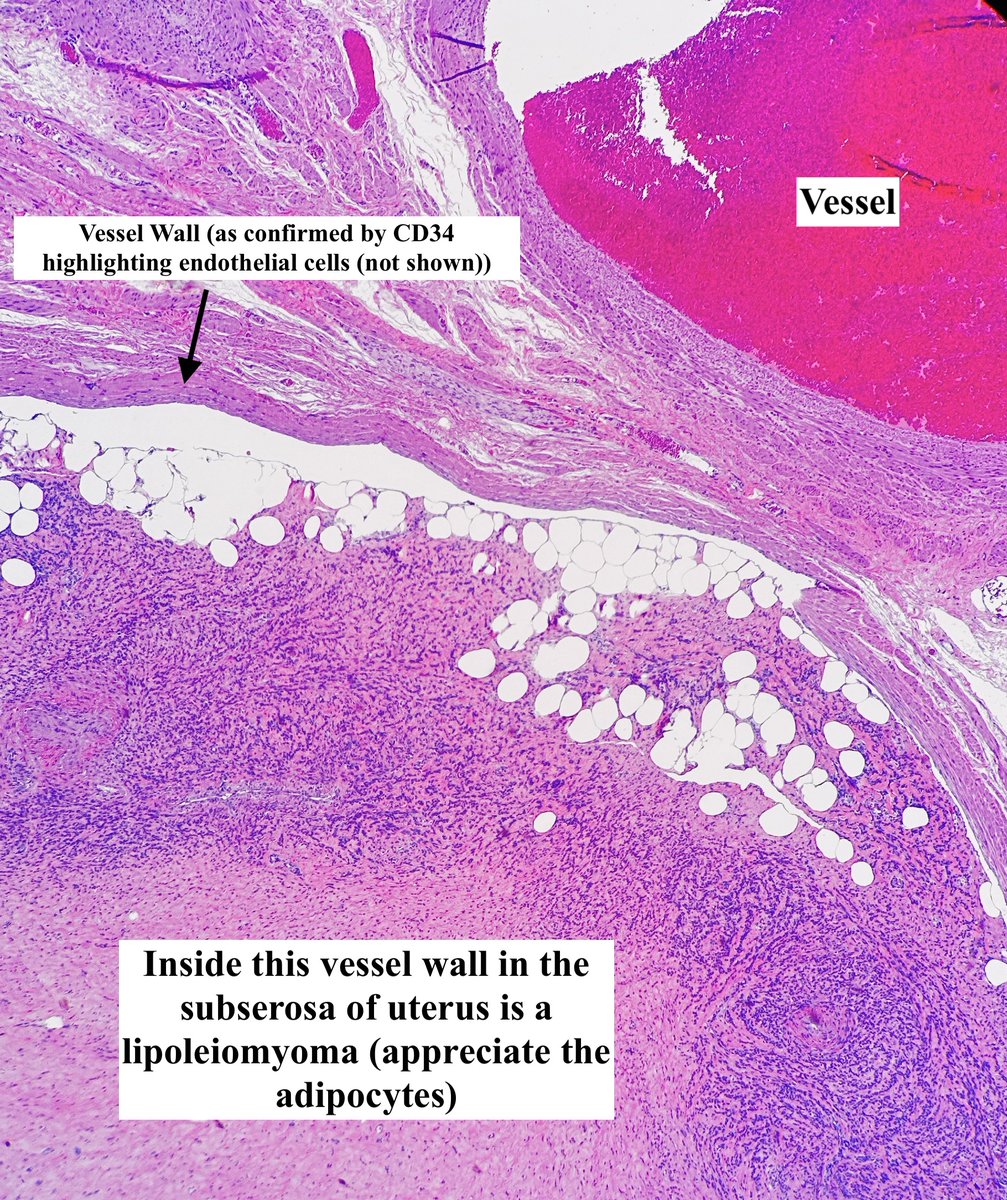
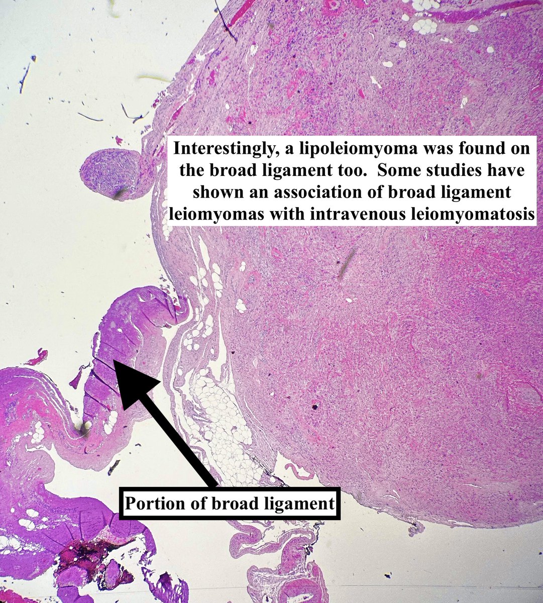
Intravenous (Lipo)Leiomyomatosis • A mouthful but basically a fibroid w fat elements inside a vessel that itself is outside a fibroid • Not staged • Extrauterine extension occurs ~ 30% of times • This case also had a broad ligament leiomyoma #pathagonia #gynpath


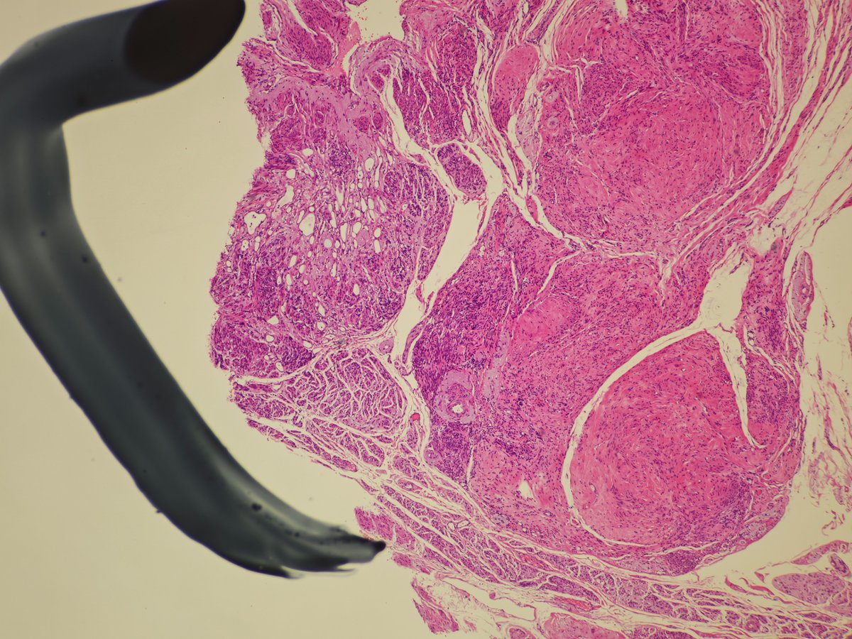
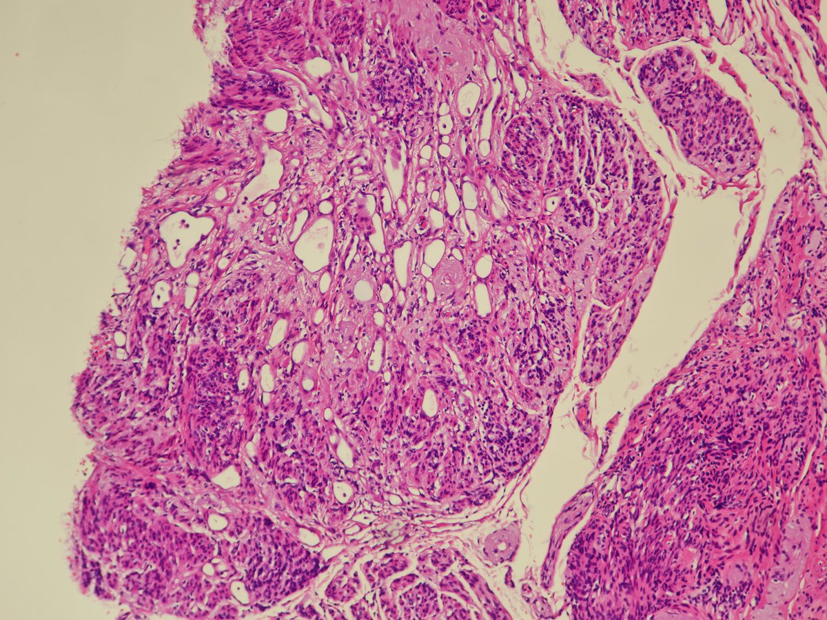
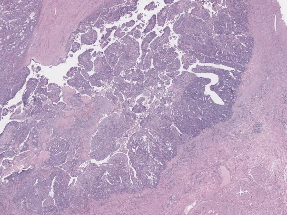
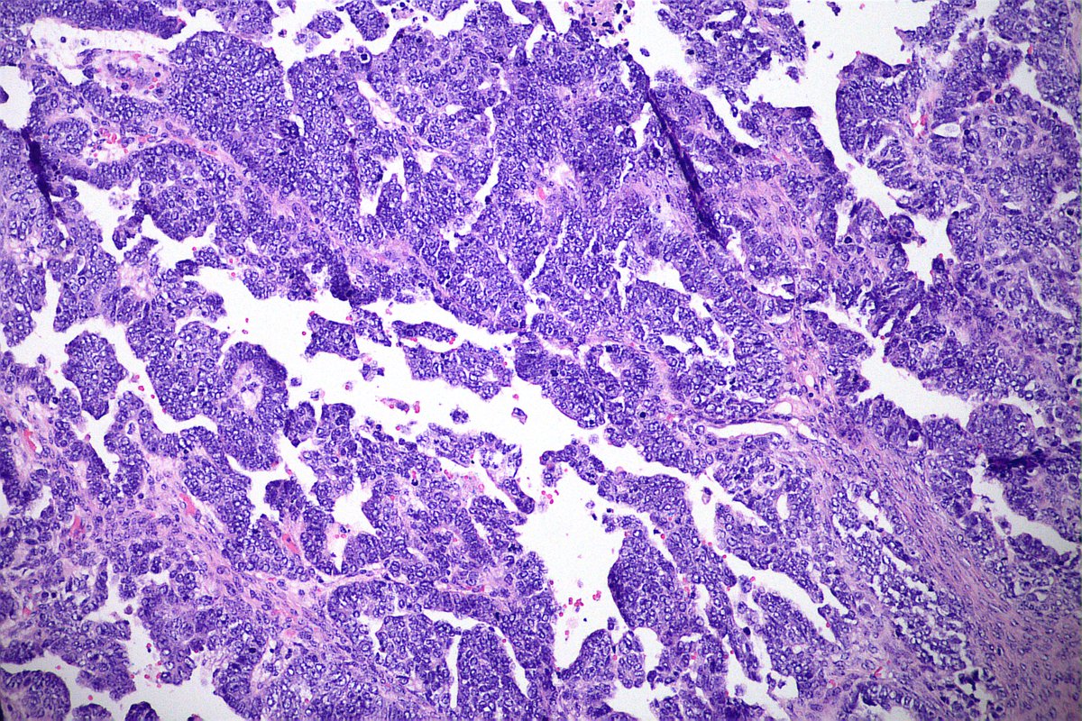
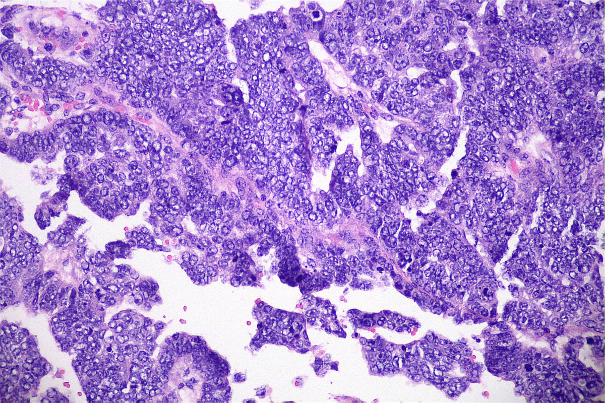
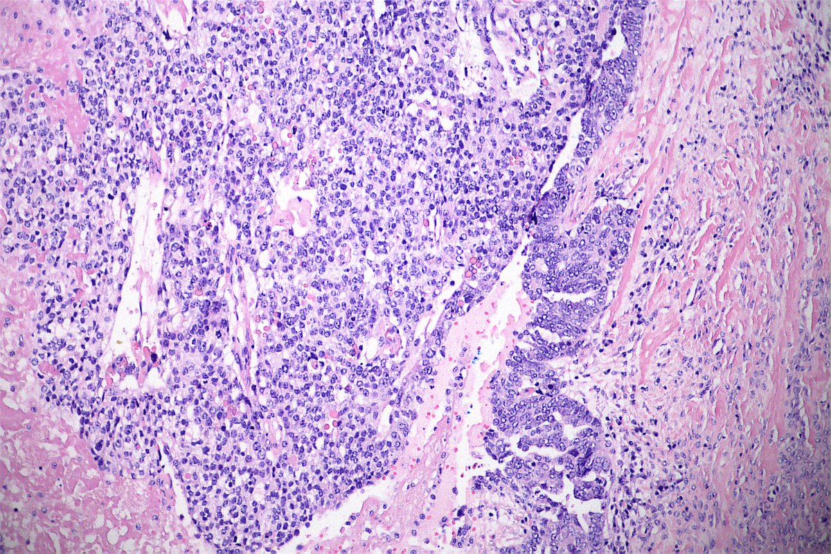
65-year-old patient with bilateral ovarian tumor and omentum implant. Almost all of the avaries with Ca serous morphology and this abrupt focus (last 2 pics). What do you think? #gynpath

🔬 Molar Pregnancy ~ Trophoblastic cell proliferation around a villous from a complete hydatidiform mole ~ #GYNpath #PediPath #Pathology
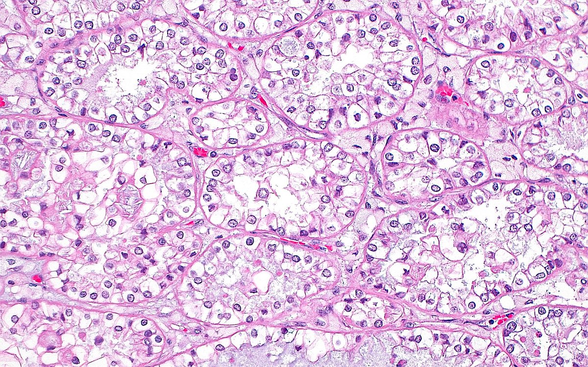
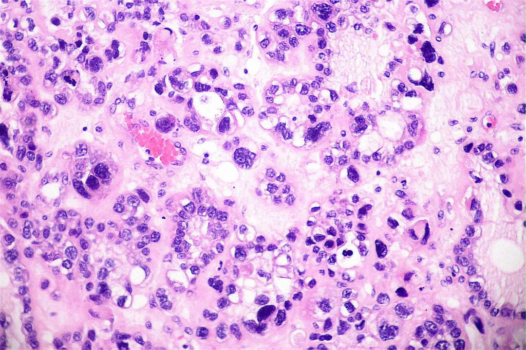
🔬Ovary: Clear cell carcinoma 👉Architectural pattern: tubulo-cystic, papillary and solid. 👉Cells with clear and eosinophilic cytoplasm. 👉Do not grade these Ca - All are high grade. 👉Nuclear pleomorphism may be present focally and mitotic activity is generally low. #gynpath

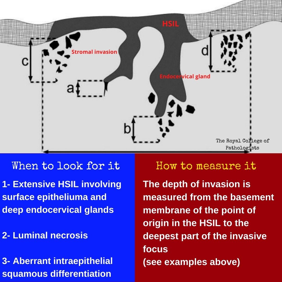
Uterine Cervix-When to look for and how to measure invasive focus. A great Saturday everyone ! #gynpath #pathology
Something went wrong.
Something went wrong.
United States Trends
- 1. #MerloFC N/A
- 2. #NIKKEAnniversary N/A
- 3. #NIKKETTStar N/A
- 4. Good Saturday N/A
- 5. #sabchella N/A
- 6. #WWEHOF N/A
- 7. Jalen Green N/A
- 8. Madonna N/A
- 9. Whale - Buy N/A
- 10. Kerr N/A
- 11. Steph N/A
- 12. Warriors N/A
- 13. Suns N/A
- 14. AJ Styles N/A
- 15. Draymond N/A
- 16. Podz N/A
- 17. Like a Prayer N/A
- 18. Scott Foster N/A
- 19. The Atlantic N/A
- 20. Thunder in 4 N/A