#scanningelectronmicroscopy search results

'Withered Plant' Scanning Electron Microscope No.684 Image width: 0.35mm Archived DOI: 10.5281/zenodo.17569069 #kikohmatsuura #witheredplant #scanningelectronmicroscopy #sciart


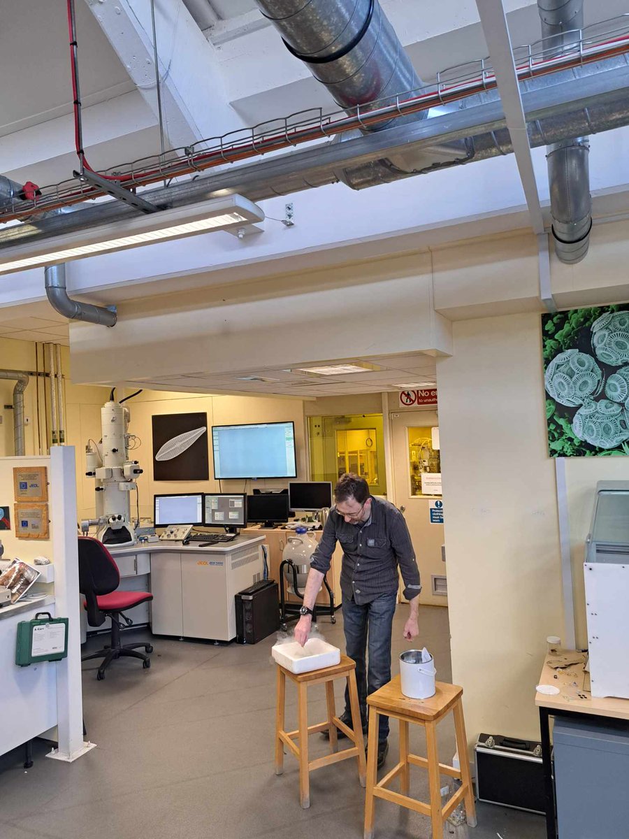
It's the greatest feeling when your testing hypothesis turns out to be right on point, and super cool to test it out with a new technique. #scanningelectronmicroscopy #finland #environmentalexchange #PhD

'Withered Plant' Scanning Electron Microscope No.683 Image width: 3mm Archived DOI: 10.5281/zenodo.17548816 #kikohmatsuura #witheredplant #scanningelectronmicroscopy #sciart

'Withered Plant' Scanning Electron Microscope No.682 Image width: 1.17mm Archived DOI: 10.5281/zenodo.17542654 #kikohmatsuura #witheredplant #scanningelectronmicroscopy #sciart


Image of the Day ~ A beautiful SEM image of Aspergillus fumigatus taken by Nurgul Daniyeva, Nazarbayev University, Kazakhstan with the JEOL JSM- IT200 SEM. #microscopy #fungus #scanningelectronmicroscopy #SEM #mold #imagecontest


Five dollar bill in a scanning electron microscope #microscopy #sem #scanningelectronmicroscopy #scanningelectronmicroscope #money #dollar


Caption This! Coated sample imaged at 3kV. #scanningelectronmicroscopy #insects #lowkV #entomology #bugs


Last year on #ValentinesDay I was doing some routine #scanningelectronmicroscopy imaging on #graphene when I found a #heart-shaped nanoparticle! Of course I had to dedicate it to my wonderful husband @Wittylama 🥰 #nerdy #cheesy #love


Dydd Gŵyl Dewi Hapus (Happy St David's Day) from the nmRC! Take a closer look at some daffodil pollen with these stunning SEM images #DyddGŵylDewiHapus #Daffodil #ScanningElectronMicroscopy @UniofNottingham @UoNScience @UoNPressOffice


6 Important Things About #ScanningElectronMicroscopy (SEM) #sme #businesstips businesspartnermagazine.com/important-thin…


SEM, EDS and XRD techniques reveal crucial info on corrosion products. Tap to learn more: bit.ly/3pkgGTr #AnalyzingCorrosionProducts #ScanningElectronMicroscopy #EnergyDispersiveXRaySpectroscopy #XRayDiffraction #VitalInformation


Spicing up the #SciArtPortfolioWeek with salt and pepper in #collaboration with @LangleyAPhoto #ScanningElectronMicroscopy #KissedByElectrons 😘 ⚡️

'Withered Plant' Scanning Electron Microscope No.681 Image width: 2.48mm Archived DOI: 10.5281/zenodo.17529240 #kikohmatsuura #witheredplant #scanningelectronmicroscopy #sciart

'Withered Plant' Scanning Electron Microscope No.680 Image width: 1.97mm Archived DOI: 10.5281/zenodo.17521658 #kikohmatsuura #witheredplant #scanningelectronmicroscopy #sciart

May #ImageContest Winner is a fully-hydrated insect sample imaged in close-to-native state. Credit: Anurag Sharma & Parviz Daniel Hejazi Pastor, The Rockefeller University. JEOL JSM-IT500HR #scanningelectronmicroscopy #insect #entomology #Cryosem #PoorMansCryo Congratulations!


🔬Are you working with #ScanningElectronMicroscopy & based near Brno, Czech Republic? We're thrilled to be part of the first edition of ICEM 2025 (In-Situ and Correlative Electron Microscopy), 🚀a must-attend conference & workshop for #ElectronMicroscopy ...

Spiderwebs 🕸️ in an electron microscope They’ve collected lots of interesting little pieces—what I found most fascinating is the scales of bugs that we can see on a few of them Evidence of a recent meal or a near miss #microscopy #scanningelectronmicroscopy #nature #science


🔥 Read our Review Paper 📚 Mineral Characterization Using Scanning Electron Microscopy (SEM): A Review of the Fundamentals, Advancements, and Research Directions 🔗 mdpi.com/2076-3417/13/2… 👨🔬 by Asif Ali et al. #scanningelectronmicroscopy #minerals


Over the past two weeks, we welcomed 10 different schools from the Devon area to visit the lab and learn all about #ElectronMicroscopy from biological sample preparation to sample analysis! 👩🔬 🔬 🦠 #TransmissionElectronMicroscopy #ScanningElectronMicroscopy #Biology #Research

'Withered Plant' Scanning Electron Microscope No.684 Image width: 0.35mm Archived DOI: 10.5281/zenodo.17569069 #kikohmatsuura #witheredplant #scanningelectronmicroscopy #sciart
'Withered Plant' Scanning Electron Microscope No.683 Image width: 3mm Archived DOI: 10.5281/zenodo.17548816 #kikohmatsuura #witheredplant #scanningelectronmicroscopy #sciart
'Withered Plant' Scanning Electron Microscope No.682 Image width: 1.17mm Archived DOI: 10.5281/zenodo.17542654 #kikohmatsuura #witheredplant #scanningelectronmicroscopy #sciart
'Withered Plant' Scanning Electron Microscope No.681 Image width: 2.48mm Archived DOI: 10.5281/zenodo.17529240 #kikohmatsuura #witheredplant #scanningelectronmicroscopy #sciart
'Withered Plant' Scanning Electron Microscope No.680 Image width: 1.97mm Archived DOI: 10.5281/zenodo.17521658 #kikohmatsuura #witheredplant #scanningelectronmicroscopy #sciart
Caption This! Coated sample imaged at 3kV. #scanningelectronmicroscopy #insects #lowkV #entomology #bugs
It's the greatest feeling when your testing hypothesis turns out to be right on point, and super cool to test it out with a new technique. #scanningelectronmicroscopy #finland #environmentalexchange #PhD
Spicing up the #SciArtPortfolioWeek with salt and pepper in #collaboration with @LangleyAPhoto #ScanningElectronMicroscopy #KissedByElectrons 😘 ⚡️
Dydd Gŵyl Dewi Hapus (Happy St David's Day) from the nmRC! Take a closer look at some daffodil pollen with these stunning SEM images #DyddGŵylDewiHapus #Daffodil #ScanningElectronMicroscopy @UniofNottingham @UoNScience @UoNPressOffice
Five dollar bill in a scanning electron microscope #microscopy #sem #scanningelectronmicroscopy #scanningelectronmicroscope #money #dollar
May #ImageContest Winner is a fully-hydrated insect sample imaged in close-to-native state. Credit: Anurag Sharma & Parviz Daniel Hejazi Pastor, The Rockefeller University. JEOL JSM-IT500HR #scanningelectronmicroscopy #insect #entomology #Cryosem #PoorMansCryo Congratulations!

🌲#Forests #HighlyAccessedPapers in 2022 Series "#ScanningElectronMicroscopy Protocol for Studying Anatomy of Highly Degraded #WaterloggedArchaeologicalWood", by Angela Balzano @AngelaBalzano5 et al. 👈 📚mdpi.com/1999-4907/13/2… ⚡️#woodanatomy #woodpreservation #protocol #EDX

Image of the Day ~ A beautiful SEM image of Aspergillus fumigatus taken by Nurgul Daniyeva, Nazarbayev University, Kazakhstan with the JEOL JSM- IT200 SEM. #microscopy #fungus #scanningelectronmicroscopy #SEM #mold #imagecontest

When things don't work in the lab and you want to run and hide somewhere.. try a multilayer cave! #scanningelectronmicroscopy #SEM #metamaterials #nanoscience

SEM, EDS and XRD techniques reveal crucial info on corrosion products. Tap to learn more: bit.ly/3pkgGTr #AnalyzingCorrosionProducts #ScanningElectronMicroscopy #EnergyDispersiveXRaySpectroscopy #XRayDiffraction #VitalInformation

Endophytic Bacteria from Lannea coromandelica for Enhancing Silver Nanoparticle Biosynthesis Read the Article here: bit.ly/3VDNOHq #EndophyticBacteria #Lanneacoromandelica #ScanningElectronMicroscopy #SilverNanoparticle #Biomedical #Pharmacology #MedicalMicrobiology

Holiday Special Image of the Day ~ "Christmas MicroTrees" Microarray of 3D-printed polymer microneedles; CREDIT: Vijayasankar Raman, University of Mississippi; SEM image from JEOL JSM-5600. #imagecontest #scanningelectronmicroscopy #polymer

Over the past two weeks, we welcomed 10 different schools from the Devon area to visit the lab and learn all about #ElectronMicroscopy from biological sample preparation to sample analysis! 👩🔬 🔬 🦠 #TransmissionElectronMicroscopy #ScanningElectronMicroscopy #Biology #Research
Last year on #ValentinesDay I was doing some routine #scanningelectronmicroscopy imaging on #graphene when I found a #heart-shaped nanoparticle! Of course I had to dedicate it to my wonderful husband @Wittylama 🥰 #nerdy #cheesy #love

Today we are exhibiting at the SEMT meeting at the Natural History Museum in London. Stop by our stand and find out more about our solutions for electron microscopy. #electronmicroscopy #scanningelectronmicroscopy #environmentalisolation #samplepreparation


Microstructural and Adsorption Behavior of Non-Polar Amino Acids in Soil Amended with Polyethylene Glycol Read the Article here: bit.ly/4eBbowX #Aminoacid #Scanningelectronmicroscopy #soilstabilization #watersolublepolymer #Xraydiffraction #chemistry #biochemistry

Read the Article here - bit.ly/3UOJtAr Studies on Minimization of Pollutants from Sugar Industry Effluent by Using a Combination of Metal Coagulant and Polymer #Chemicaloxygendemand #Scanningelectronmicroscopy #Turbidity #Totalsuspendedsolids #Totaldissolvedsolids

Spiderwebs 🕸️ in an electron microscope They’ve collected lots of interesting little pieces—what I found most fascinating is the scales of bugs that we can see on a few of them Evidence of a recent meal or a near miss #microscopy #scanningelectronmicroscopy #nature #science

Christmas Cheer Spreads Worldwide as Overseas Customers Receive "Christmas Presents" - SEM3200 in Saudi Arabia and SEM5000 Pro in South Korea. Learn more:buff.ly/407aC5C #ElectronMicroscope#CIQTEK #SEMmicroscope #scanningelectronmicroscopy #GSEM


An Editors' Pick via #OPG_OpEx: Subwavelength-modulated silicon photonics for low-energy free-electron-photon interactions bit.ly/3AshWPj #DielectricWaveguides #ScanningElectronMicroscopy @ucdavis

Something went wrong.
Something went wrong.
United States Trends
- 1. Daboll 35.9K posts
- 2. Pond 237K posts
- 3. Schoen 18.4K posts
- 4. Schoen 18.4K posts
- 5. Veterans Day 21.2K posts
- 6. Joe Burrow 5,585 posts
- 7. Giants 71.4K posts
- 8. Go Birds 11.4K posts
- 9. Dart 23.2K posts
- 10. Kim Davis 12.6K posts
- 11. #ROBOGIVE 1,056 posts
- 12. Marines 60.5K posts
- 13. Joe Dirt N/A
- 14. Zendaya 7,913 posts
- 15. Johnny Carson N/A
- 16. Jeffries 40.2K posts
- 17. #jimromeonx N/A
- 18. Hanoi Jane N/A
- 19. Semper Fi 11.6K posts
- 20. Kafka 9,501 posts