#scanningelectronmicroscopy search results
'Withered Plant' Scanning Electron Microscope No.684 Image width: 0.35mm Archived DOI: 10.5281/zenodo.17569069 #kikohmatsuura #witheredplant #scanningelectronmicroscopy #sciart

It's the greatest feeling when your testing hypothesis turns out to be right on point, and super cool to test it out with a new technique. #scanningelectronmicroscopy #finland #environmentalexchange #PhD

'Withered Plant' Scanning Electron Microscope No.683 Image width: 3mm Archived DOI: 10.5281/zenodo.17548816 #kikohmatsuura #witheredplant #scanningelectronmicroscopy #sciart

'Withered Plant' Scanning Electron Microscope No.681 Image width: 2.48mm Archived DOI: 10.5281/zenodo.17529240 #kikohmatsuura #witheredplant #scanningelectronmicroscopy #sciart

'Withered Plant' Scanning Electron Microscope No.682 Image width: 1.17mm Archived DOI: 10.5281/zenodo.17542654 #kikohmatsuura #witheredplant #scanningelectronmicroscopy #sciart

'Withered Plant' Scanning Electron Microscope No.680 Image width: 1.97mm Archived DOI: 10.5281/zenodo.17521658 #kikohmatsuura #witheredplant #scanningelectronmicroscopy #sciart

Caption This! Coated sample imaged at 3kV. #scanningelectronmicroscopy #insects #lowkV #entomology #bugs

Five dollar bill in a scanning electron microscope #microscopy #sem #scanningelectronmicroscopy #scanningelectronmicroscope #money #dollar


Last year on #ValentinesDay I was doing some routine #scanningelectronmicroscopy imaging on #graphene when I found a #heart-shaped nanoparticle! Of course I had to dedicate it to my wonderful husband @Wittylama 🥰 #nerdy #cheesy #love

Dydd Gŵyl Dewi Hapus (Happy St David's Day) from the nmRC! Take a closer look at some daffodil pollen with these stunning SEM images #DyddGŵylDewiHapus #Daffodil #ScanningElectronMicroscopy @UniofNottingham @UoNScience @UoNPressOffice

SEM, EDS and XRD techniques reveal crucial info on corrosion products. Tap to learn more: bit.ly/3pkgGTr #AnalyzingCorrosionProducts #ScanningElectronMicroscopy #EnergyDispersiveXRaySpectroscopy #XRayDiffraction #VitalInformation

Spicing up the #SciArtPortfolioWeek with salt and pepper in #collaboration with @LangleyAPhoto #ScanningElectronMicroscopy #KissedByElectrons 😘 ⚡️

6 Important Things About #ScanningElectronMicroscopy (SEM) #sme #businesstips businesspartnermagazine.com/important-thin…
May #ImageContest Winner is a fully-hydrated insect sample imaged in close-to-native state. Credit: Anurag Sharma & Parviz Daniel Hejazi Pastor, The Rockefeller University. JEOL JSM-IT500HR #scanningelectronmicroscopy #insect #entomology #Cryosem #PoorMansCryo Congratulations!

🔬Are you working with #ScanningElectronMicroscopy & based near Brno, Czech Republic? We're thrilled to be part of the first edition of ICEM 2025 (In-Situ and Correlative Electron Microscopy), 🚀a must-attend conference & workshop for #ElectronMicroscopy ...

Spiderwebs 🕸️ in an electron microscope They’ve collected lots of interesting little pieces—what I found most fascinating is the scales of bugs that we can see on a few of them Evidence of a recent meal or a near miss #microscopy #scanningelectronmicroscopy #nature #science


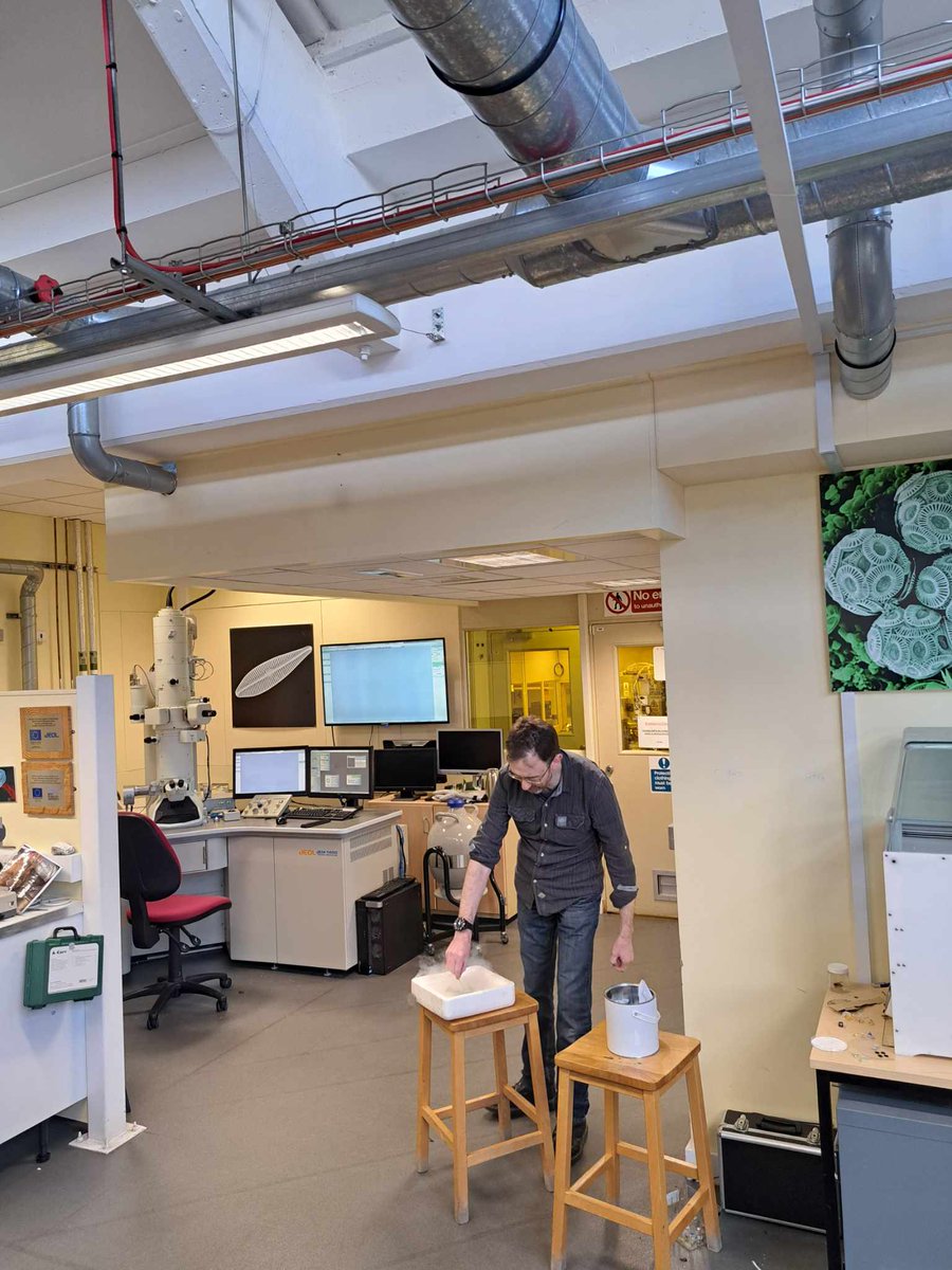
Over the past two weeks, we welcomed 10 different schools from the Devon area to visit the lab and learn all about #ElectronMicroscopy from biological sample preparation to sample analysis! 👩🔬 🔬 🦠 #TransmissionElectronMicroscopy #ScanningElectronMicroscopy #Biology #Research


🔥 Read our Review Paper 📚 Mineral Characterization Using Scanning Electron Microscopy (SEM): A Review of the Fundamentals, Advancements, and Research Directions 🔗 mdpi.com/2076-3417/13/2… 👨🔬 by Asif Ali et al. #scanningelectronmicroscopy #minerals

Image of the Day ~ A beautiful SEM image of Aspergillus fumigatus taken by Nurgul Daniyeva, Nazarbayev University, Kazakhstan with the JEOL JSM- IT200 SEM. #microscopy #fungus #scanningelectronmicroscopy #SEM #mold #imagecontest

'Withered Plant' Scanning Electron Microscope No.684 Image width: 0.35mm Archived DOI: 10.5281/zenodo.17569069 #kikohmatsuura #witheredplant #scanningelectronmicroscopy #sciart

'Withered Plant' Scanning Electron Microscope No.683 Image width: 3mm Archived DOI: 10.5281/zenodo.17548816 #kikohmatsuura #witheredplant #scanningelectronmicroscopy #sciart

'Withered Plant' Scanning Electron Microscope No.682 Image width: 1.17mm Archived DOI: 10.5281/zenodo.17542654 #kikohmatsuura #witheredplant #scanningelectronmicroscopy #sciart

'Withered Plant' Scanning Electron Microscope No.681 Image width: 2.48mm Archived DOI: 10.5281/zenodo.17529240 #kikohmatsuura #witheredplant #scanningelectronmicroscopy #sciart

'Withered Plant' Scanning Electron Microscope No.680 Image width: 1.97mm Archived DOI: 10.5281/zenodo.17521658 #kikohmatsuura #witheredplant #scanningelectronmicroscopy #sciart

Caption This! Coated sample imaged at 3kV. #scanningelectronmicroscopy #insects #lowkV #entomology #bugs

It's the greatest feeling when your testing hypothesis turns out to be right on point, and super cool to test it out with a new technique. #scanningelectronmicroscopy #finland #environmentalexchange #PhD

Image of the Day ~ A beautiful SEM image of Aspergillus fumigatus taken by Nurgul Daniyeva, Nazarbayev University, Kazakhstan with the JEOL JSM- IT200 SEM. #microscopy #fungus #scanningelectronmicroscopy #SEM #mold #imagecontest

Five dollar bill in a scanning electron microscope #microscopy #sem #scanningelectronmicroscopy #scanningelectronmicroscope #money #dollar


Dydd Gŵyl Dewi Hapus (Happy St David's Day) from the nmRC! Take a closer look at some daffodil pollen with these stunning SEM images #DyddGŵylDewiHapus #Daffodil #ScanningElectronMicroscopy @UniofNottingham @UoNScience @UoNPressOffice

Spicing up the #SciArtPortfolioWeek with salt and pepper in #collaboration with @LangleyAPhoto #ScanningElectronMicroscopy #KissedByElectrons 😘 ⚡️

When things don't work in the lab and you want to run and hide somewhere.. try a multilayer cave! #scanningelectronmicroscopy #SEM #metamaterials #nanoscience

May #ImageContest Winner is a fully-hydrated insect sample imaged in close-to-native state. Credit: Anurag Sharma & Parviz Daniel Hejazi Pastor, The Rockefeller University. JEOL JSM-IT500HR #scanningelectronmicroscopy #insect #entomology #Cryosem #PoorMansCryo Congratulations!

Today we are exhibiting at the SEMT meeting at the Natural History Museum in London. Stop by our stand and find out more about our solutions for electron microscopy. #electronmicroscopy #scanningelectronmicroscopy #environmentalisolation #samplepreparation

🌲#Forests #HighlyAccessedPapers in 2022 Series "#ScanningElectronMicroscopy Protocol for Studying Anatomy of Highly Degraded #WaterloggedArchaeologicalWood", by Angela Balzano @AngelaBalzano5 et al. 👈 📚mdpi.com/1999-4907/13/2… ⚡️#woodanatomy #woodpreservation #protocol #EDX

Over the past two weeks, we welcomed 10 different schools from the Devon area to visit the lab and learn all about #ElectronMicroscopy from biological sample preparation to sample analysis! 👩🔬 🔬 🦠 #TransmissionElectronMicroscopy #ScanningElectronMicroscopy #Biology #Research


Holiday Special Image of the Day ~ "Christmas MicroTrees" Microarray of 3D-printed polymer microneedles; CREDIT: Vijayasankar Raman, University of Mississippi; SEM image from JEOL JSM-5600. #imagecontest #scanningelectronmicroscopy #polymer

Christmas Cheer Spreads Worldwide as Overseas Customers Receive "Christmas Presents" - SEM3200 in Saudi Arabia and SEM5000 Pro in South Korea. Learn more:buff.ly/407aC5C #ElectronMicroscope#CIQTEK #SEMmicroscope #scanningelectronmicroscopy #GSEM


Last year on #ValentinesDay I was doing some routine #scanningelectronmicroscopy imaging on #graphene when I found a #heart-shaped nanoparticle! Of course I had to dedicate it to my wonderful husband @Wittylama 🥰 #nerdy #cheesy #love

SEM, EDS and XRD techniques reveal crucial info on corrosion products. Tap to learn more: bit.ly/3pkgGTr #AnalyzingCorrosionProducts #ScanningElectronMicroscopy #EnergyDispersiveXRaySpectroscopy #XRayDiffraction #VitalInformation

The lack of the summer sun 🌥 in the UK hasn't stopped us from heading to the beach! For #MicroscopyMonday, here's a #ScanningElectronMicroscopy image of sand from nearby Bovisand ⛱ #ElectronMicroscopy #Sand #Beach #Summer #Imaging #Geology #Science @JEOLEUROPE

Microstructural and Adsorption Behavior of Non-Polar Amino Acids in Soil Amended with Polyethylene Glycol Read the Article here: bit.ly/4eBbowX #Aminoacid #Scanningelectronmicroscopy #soilstabilization #watersolublepolymer #Xraydiffraction #chemistry #biochemistry

Metal fractures can occur in various failure modes. CIQTEK scanning electron microscope characterization can help researchers quickly analyze fracture surfaces. Learn more: buff.ly/3Y3Ta0Q #CIQTEK #SEMmicroscope #scanningelectronmicroscopy #electronmicroscopy


Spiderwebs 🕸️ in an electron microscope They’ve collected lots of interesting little pieces—what I found most fascinating is the scales of bugs that we can see on a few of them Evidence of a recent meal or a near miss #microscopy #scanningelectronmicroscopy #nature #science


Part4(buff.ly/48LXu8x ): Improvement of low-frequency vibration environment Improving low-frequency vibrations can be costly, and sometimes it is not feasible due to environmental constraints. #CIQTEK #SEMmicroscope #scanningelectronmicroscopy #electronmicroscopy


Something went wrong.
Something went wrong.
United States Trends
- 1. Pond 218K posts
- 2. Kim Davis 3,754 posts
- 3. #IDontWantToOverreactBUT N/A
- 4. $BNKK 1,070 posts
- 5. Go Birds 6,137 posts
- 6. Semper Fi 7,351 posts
- 7. Happy 250th 9,541 posts
- 8. #MondayMotivation 40.9K posts
- 9. $LMT $450.50 Lockheed F-35 N/A
- 10. #MYNZ N/A
- 11. $SENS $0.70 Senseonics CGM N/A
- 12. $APDN $0.20 Applied DNA N/A
- 13. Obergefell 2,624 posts
- 14. Good Monday 47.9K posts
- 15. Obamacare 22.9K posts
- 16. Edmund Fitzgerald 6,016 posts
- 17. Victory Monday 3,076 posts
- 18. Veterans Day 21.6K posts
- 19. #USMC 1,454 posts
- 20. Rudy Giuliani 31.2K posts


