#smartspim search results

Dr. @kramadi's lab at NYU Abu Dhabi used the LifeCanvas pipeline to develop CORAL, an ingestible device to non-invasively sample the small intestine #microbiome. The #SmartSPIM video below shows blood vessels (pink) & epithelial cells (blue) in rat duodenum.

Happy #microscopymonday 🎉 Mouse kidney captured with our new #SmartSPIM @LifeCanvasTech 🔬 Thank you @Carolinar353 for providing this beautiful sample #SciArt #bioart #microscopy

The most common cause of surgical treatment for childhood #epilepsy is a brain malformation called focal cortical dysplasia (FCD). At @CUAnschutz, Paige Hoffman uses #SmartSPIM whole-brain data to study the mechanisms that lead to epilepsy in FCD.
This mouse brain (5XFAD x BXD29) was stained using SmartBatch+ with Thioflavin S dye, which binds to Beta Amyloid plaques and Tau tangles. It was captured with #SmartSPIM at 3.6X magnification. Sample courtesy of David Ashbrook & Robert W. Williams, UTHSC; G. Allan Johnson, Duke.
The intact mouse brain was labeled with NeuN using SmartBatch+ and imaged with #SmartSPIM at 3.6X magnification. The images were aligned to the Allen Mouse Brain Atlas using an automated, multi-stage registration pipeline. Sample courtesy of Ian Meng, University of New England.
The neuropeptide Orexin is produced by neurons in the hypothalamus and regulates sleep, appetite, and reward-seeking behaviors. Orexin blockers are being studied as a treatment for addiction and sleep disorders. Mouse brain labeled with SmartBatch+ and imaged with #SmartSPIM.
Let me introduce you to our newest super fast #lightsheet microscope 🎉 #SmartSPIM from @LifeCanvasTech 🤩🔬#microscopy


Why are half of people with Down syndrome born with heart defects? That’s what @SanjeevRanade & Feiya Li et al. uncovered in a new study using single-cell #transcriptomics + #SmartSPIM imaging to characterize the impact of trisomy 21 on heart development: hubs.li/Q03QLB440

Beta Tubulin (red) labels type I spiral ganglion neurons in the mouse cochlea. NF-H (blue) labels the auditory nerve fibers, which carry information from the hair cells to the cochlear nucleus. Imaged with #SmartSPIM at 9X. Sample courtesy of Dr. Bohua Hu, UT Dallas.
#FluorescenceFriday: #SmartSPIM video from the Pattabiraman Lab (@Yale)! #GFP (green) & #TranssynapticTracing (red) reveal prefrontal cortex & thalamus connectivity in a neonatal mouse brain. Full video & details: bit.ly/3UUwKx0 Thanks to @kartikpattabir1 @_dan_doyle
Thy1-GFP transgenic mouse lines are used to generate stunning images of fine neuronal structures. Different Thy1 mouse lines express GFP across variable cell populations, hitting around 10% of cells in a particular group. Clear+ delipidation and #SmartSPIM imaging at 3.6X.
These Purkinje cells tagged with mOrange2 are showing off their distinctive fan shape and single plane orientation. Purkinje cells are found only in the cerebellar cortex and are involved in motor control. #SmartSPIM, 9X magnification. Sample courtesy of Michelle Antoine, NIH.
DeltaFosB expression in a mouse brain, labeled using LifeCanvas protocols and imaged with #SmartSPIM at 3.6X magnification. Sample courtesy of Joselyn Soto, UCLA.
Today for #FluorescenceFriday we look at C-FOS labeling and cell detection in mouse brain imaged with our #SmartSPIM light sheet microscope. Researching the complexities of brain activity? Learn more about how we can help: bit.ly/4aiXQng
GFAP labeling astrocytes in the corpus callosum and cortex in a mouse brain. The whole mouse brain was cleared with Clear+ delipidation buffer, labeled with SmartBatch+, and imaged with #SmartSPIM at 3.6X magnification. #tissueclearing #neuroscience #brainresearch
#FluorescenceFriday Check out this stunning video of GFAP-labeled astrocytes in a mouse brain, imaged with #SmartSPIM at 15X magnification! Contact us to learn more: lifecanvastech.com/contact-us/ #Brain #Neuroscience #Research
🏆Congrats to our #LifeCanvasTravelGrant Winners for the @SfNtweets 2024 meeting. Their research using our advanced imaging tech including #SmartSPIM, #SmartBatch+, #Clear+ clearing pipeline, stood out brilliantly. @ShreyasMSurya @albit_caban and Samuel Reynolds! Stay tuned!

The more, the merrier ! Fast light-sheet imaging of four brains at once 🧠 #lightsheet #SmartSPIM @LifeCanvasTech

Check it out: β-amyloid plaques + astrocytes in an AD model mouse brain from @Taconic Biosciences, imaged with #SmartSPIM! #SmartSPIM provides accuracy, speed, and flexibility for intact tissues. Talk to our experts: bit.ly/3LiDX4B #research #neuroscience #microscopy
New research! @UF Brain Tumor Immunotherapy Program developed a novel immunotherapy for GBM using AAV gene therapy & anti-PD-1. With #SmartSPIM 3D light sheet imaging, they visualized the tumor landscape. Read full article: go.nature.com/3LEvitA #imaging #research

Dr. @kramadi's lab at NYU Abu Dhabi used the LifeCanvas pipeline to develop CORAL, an ingestible device to non-invasively sample the small intestine #microbiome. The #SmartSPIM video below shows blood vessels (pink) & epithelial cells (blue) in rat duodenum.
Why are half of people with Down syndrome born with heart defects? That’s what @SanjeevRanade & Feiya Li et al. uncovered in a new study using single-cell #transcriptomics + #SmartSPIM imaging to characterize the impact of trisomy 21 on heart development: hubs.li/Q03QLB440
The most common cause of surgical treatment for childhood #epilepsy is a brain malformation called focal cortical dysplasia (FCD). At @CUAnschutz, Paige Hoffman uses #SmartSPIM whole-brain data to study the mechanisms that lead to epilepsy in FCD.
The intact mouse brain was labeled with NeuN using SmartBatch+ and imaged with #SmartSPIM at 3.6X magnification. The images were aligned to the Allen Mouse Brain Atlas using an automated, multi-stage registration pipeline. Sample courtesy of Ian Meng, University of New England.
This mouse brain (5XFAD x BXD29) was stained using SmartBatch+ with Thioflavin S dye, which binds to Beta Amyloid plaques and Tau tangles. It was captured with #SmartSPIM at 3.6X magnification. Sample courtesy of David Ashbrook & Robert W. Williams, UTHSC; G. Allan Johnson, Duke.
The neuropeptide Orexin is produced by neurons in the hypothalamus and regulates sleep, appetite, and reward-seeking behaviors. Orexin blockers are being studied as a treatment for addiction and sleep disorders. Mouse brain labeled with SmartBatch+ and imaged with #SmartSPIM.
Beta Tubulin (red) labels type I spiral ganglion neurons in the mouse cochlea. NF-H (blue) labels the auditory nerve fibers, which carry information from the hair cells to the cochlear nucleus. Imaged with #SmartSPIM at 9X. Sample courtesy of Dr. Bohua Hu, UT Dallas.
Thy1-GFP transgenic mouse lines are used to generate stunning images of fine neuronal structures. Different Thy1 mouse lines express GFP across variable cell populations, hitting around 10% of cells in a particular group. Clear+ delipidation and #SmartSPIM imaging at 3.6X.
GFAP labeling astrocytes in the corpus callosum and cortex in a mouse brain. The whole mouse brain was cleared with Clear+ delipidation buffer, labeled with SmartBatch+, and imaged with #SmartSPIM at 3.6X magnification. #tissueclearing #neuroscience #brainresearch
These Purkinje cells tagged with mOrange2 are showing off their distinctive fan shape and single plane orientation. Purkinje cells are found only in the cerebellar cortex and are involved in motor control. #SmartSPIM, 9X magnification. Sample courtesy of Michelle Antoine, NIH.
DeltaFosB expression in a mouse brain, labeled using LifeCanvas protocols and imaged with #SmartSPIM at 3.6X magnification. Sample courtesy of Joselyn Soto, UCLA.
The more, the merrier ! Fast light-sheet imaging of four brains at once 🧠 #lightsheet #SmartSPIM @LifeCanvasTech
Dr. Andy Stone, Manager at @BrandeisU Light Microscopy Core, calls #SmartSPIM & #SmartBatch+ game-changers after a recent demo. Ease of use & reproducibility are just the beginning! Read more: bit.ly/3YZqF3m @andystone93 @BrandeisImaging #microscopy #Neuroscience
#FluorescenceFriday Check out this stunning video of GFAP-labeled astrocytes in a mouse brain, imaged with #SmartSPIM at 15X magnification! Contact us to learn more: lifecanvastech.com/contact-us/ #Brain #Neuroscience #Research
#FluorescenceFriday: #SmartSPIM video from the Pattabiraman Lab (@Yale)! #GFP (green) & #TranssynapticTracing (red) reveal prefrontal cortex & thalamus connectivity in a neonatal mouse brain. Full video & details: bit.ly/3UUwKx0 Thanks to @kartikpattabir1 @_dan_doyle
Excited to share new research from @UCLA using ΔFosB mapping to study neural activity in OCD mouse models! They utilized our #SHIELD, #Clear+ protocols, and #SmartSPIM for 3D imaging and analysis of the striatum and cortex. #Neuroscience #Imaging bit.ly/47c3Ihf
cell.com
Astrocyte Gi-GPCR signaling corrects compulsive-like grooming and anxiety-related behaviors in...
Soto et al. used multiple approaches to assess whether and how function is affected following astrocyte Gi-DREADD activation in a mouse model of compulsive and anxiety-related behaviors that are...
🔬New study alert on brain connectivity and network changes! Oluwarotimi Folorunso's latest research used our #SHIELD, #SmartBatch+, #SmartSPIM, & #cFos analysis pipeline to map neuron activation in WT & SR-/- mice. #brainresearch #Neuroscience bit.ly/4dqpK1w

New research! @UF Brain Tumor Immunotherapy Program developed a novel immunotherapy for GBM using AAV gene therapy & anti-PD-1. With #SmartSPIM 3D light sheet imaging, they visualized the tumor landscape. Read full article: go.nature.com/3LEvitA #imaging #research
Check it out: β-amyloid plaques + astrocytes in an AD model mouse brain from @Taconic Biosciences, imaged with #SmartSPIM! #SmartSPIM provides accuracy, speed, and flexibility for intact tissues. Talk to our experts: bit.ly/3LiDX4B #research #neuroscience #microscopy
🏆Congrats to our #LifeCanvasTravelGrant Winners for the @SfNtweets 2024 meeting. Their research using our advanced imaging tech including #SmartSPIM, #SmartBatch+, #Clear+ clearing pipeline, stood out brilliantly. @ShreyasMSurya @albit_caban and Samuel Reynolds! Stay tuned!
Happy #microscopymonday 🎉 Mouse kidney captured with our new #SmartSPIM @LifeCanvasTech 🔬 Thank you @Carolinar353 for providing this beautiful sample #SciArt #bioart #microscopy
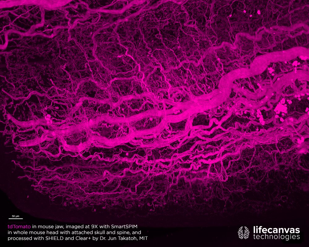
tdTomato traces innervation through this whole mouse head with attached skull and spine, processed by Dr. @jun_takatoh at MIT and imaged with #SmartSPIM in 2 hours. Our tissue clearing techniques preserve endogenous fluorescence, generating detailed 3D data. #FluorescenceFriday


Neuropeptide Y (NPY) & Somatostatin (SST) are visualized throughout this whole mouse brain, stained with SmartBatch+ and imaged with #SmartSPIM. NPY & SST are widely-expressed neuropeptides that can help identify different subtypes of GABAergic neurons. #FluorescenceFriday

Can you believe these #FluorescenceFriday #SmartSPIM images all come from the same sample? At 9X, you can see fine details like neuromuscular junctions in a whole mouse head with attached skull and spine. Sample was processed with our techniques by Dr. @jun_takatoh @mcgovernmit.


Congrats to @DrMBede et al. on making the cover of JCN with this stunning #SmartSPIM image showing MC3R expression in a whole mouse brain! #neuroscience #microscopy

Today's #FluorescenceFriday comes from @yongsookimlab: oxytocin (magenta) and vasopressin (green) in mouse brain, imaged with #SmartSPIM. To learn more about his #neuroscience research, check out the interview on our blog!

Let me introduce you to our newest super fast #lightsheet microscope 🎉 #SmartSPIM from @LifeCanvasTech 🤩🔬#microscopy
🏆Congrats to our #LifeCanvasTravelGrant Winners for the @SfNtweets 2024 meeting. Their research using our advanced imaging tech including #SmartSPIM, #SmartBatch+, #Clear+ clearing pipeline, stood out brilliantly. @ShreyasMSurya @albit_caban and Samuel Reynolds! Stay tuned!
Happy #FluorescenceFriday! Check out this stunning #SmartSPIM 15X image of astrocytes (GFAP, magenta) clustered around Beta-Amyloid plaques (cyan) in the brain of a @Taconic ARTE10 mouse model for Alzheimer's.

This #MicroscopyMonday, register to learn about innovative applications of intact #tissueclearing and #SmartSPIM imaging to the study of neurodegenerative disease! @Taconic bit.ly/3w15Ozo

Sometimes the abyss stares back. The abyss, in this case, is a mouse duodenum with vasculature (lectin) in red and Olfm4 (stem cell marker) in cyan. Sample courtesy of the Ferrara Lab at @IcahnMountSinai. #MicroscopyMonday #SmartSPIM

Action shot of our Director of Imaging Dr. Jeff Stirman talking #SmartSPIM light sheet imaging at @LSFMMBL!

Come check out #SmartSPIM and SmartBatch+ at Booth 1316 and speak to us about applications for your #neuroscience research area! #SfN22


Some action shots from our first in-house workshop! Top left to bottom right: Dr. Hsuan Lee demos #SmartSPIM acquisition, Zach Woods explains principles behind our technology, Aparna Nambiar demos SmartBatch+, and Dr. Brian Nguyen shows cleared and index-matched samples.


🔬Wrapping up an amazing week at Harvard Center for Biological Imaging! Grateful for the opportunity to showcase our #SmartSPIM, #SmartAnalytics, and #DataStorage in the Tissue Clearing Workshop. Thanks @Harvard! 🙌🔍 #Innovation #neuroscience #research #microscopy #3Dimaging


This #MicroscopyMonday, we're excited to share a conversation with imaging scientist Dr. Mike Taormina on the past, present, and future of whole brain mapping at the @AllenInstitute – and how #SmartSPIM fits into the big picture: bit.ly/41d13jB

High-quality data begins with the right tools. Try out our tissue processing techniques in your lab, with expert technical support to help tailor protocols to your application. Plus, get free #SmartSPIM imaging service for an intact sample! Sign up at: bit.ly/3Hd6t6a

Our summer workshop is happening in just 1 week! We'll have #SmartSPIM & SmartBatch+ demos, a talk by @dheerajroy7, free food, and an amazing scientific community. Register at bit.ly/3O3nNLM!

We now offer free #SmartSPIM imaging service as part of our free trial! Preserve, clear, & index-match intact samples with our kit, then send them to us to receive high-resolution 3D data. Sign up at: bit.ly/3yXhKna - Happy #FluorescenceFriday!

Have you read our interview with @AllenInstitute researcher Dr. Mike Taormina? Check out our blog to learn how #SmartSPIM light sheet microscopy is streamlining and expanding possibilities in #neuroscience research: bit.ly/41d13jB #FluorescenceFriday

Something went wrong.
Something went wrong.
United States Trends
- 1. #SmackDown 39.1K posts
- 2. #BUNCHITA N/A
- 3. Giulia 12.9K posts
- 4. #BostonBlue 4,039 posts
- 5. Caleb Wilson 4,859 posts
- 6. #OPLive 1,824 posts
- 7. Supreme Court 173K posts
- 8. Rockets 19.7K posts
- 9. Tulane 3,021 posts
- 10. #TheLastDriveIn 2,483 posts
- 11. Northwestern 4,436 posts
- 12. Podz 1,654 posts
- 13. Lash Legend 5,320 posts
- 14. Justice Jackson 3,623 posts
- 15. Chelsea Green 5,567 posts
- 16. NBA Cup 9,018 posts
- 17. Harrison Barnes N/A
- 18. Reed 23.8K posts
- 19. Justice Ketanji Brown Jackson 2,198 posts
- 20. SCOTUS 22.7K posts