#cellsegmentation search results

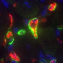
🎯 Precise #CellSegmentation is challenging. @vizgen_inc uses cell boundary stains and robust algorithms for accuracy. See these #DataImages of FFPE mouse breast tumor by researchers at @NeuroAlc, showcasing cell boundary staining and segmentation revealing natural cell shapes.
#Baysor #CellSegmentation for 2D 3D image-based #SpatialTranscriptomics Based on Transcript composition #MarkovRandomField -/+Stain image #PriorSegmentationConfidence Work with #MERFISH #smFISH #STARmap #InSituSequencing ⏫Detection of #EndothelialCell #MuralCell (Fig 5/6)…

Once we have a stitched image, how do we discover single cell phenotypes that interest us? ➡️ SPARCSpy 🔵 SPARCSpy performs precise cell segmentation, generating single-cell image datasets for all cells in a sample #cellsegmentation #singlecellimagedataset

Customize your #CellSegmentation with the new #Cellpose2 Plugin for the #Vizgen Post-Processing Tool! Visit our website to learn how you can improve segmentation results for challenging tissues with the plugin for #VPT: hubs.ly/Q02k2S000 #MERSCOPE #MERFISH #Spatialomics
🍬Treat yourself this Halloween with the new #Vizgen Post-Processing Tool for Cell Segmentation! #VPT uses #CellSegmentation methods to draw cell boundaries & generates #SingleCell output by combining the results with #MERFISH data. Learn more: hubs.ly/Q026kTq70 #MERSCOPE
In October '21, researchers from @Harvard, @BostonChildrens, & @uni_copenhagen published a paper in @NatureBiotech describing a new method for #CellSegmentation called #Baysor. Read & learn how they evaluated it with #MERFISH! hubs.ly/Q013pM8Q0 #ThrowbackThursday


Get a closer look at the superior cell segmentation and transcript information you can obtain from CosMx SMI data during our upcoming Spatial Informatics 101 Webinar on July 11. Register to attend: bit.ly/3pxrlQ4 #SpatialInformatics #CellSegmentation

What to do when #cellsegmentation does not properly work and creates an artificial #cellmask? One can always make something else out of it😊

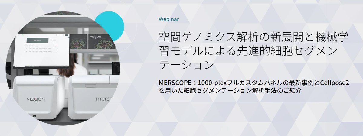
💻Webinar Alert for #SpatialTranscriptomics Customers in Japan! Developments in Spatial Genomics Analysis and Advanced Cell Segmentation 📅 Wednesday, April 24, 2024 ⏰15:00 - 15:45 JST 🎙️Wenbin Gu, Primetech Co. 🔗 hubs.ly/Q02sQSrr0 #CellSegmentation #MERSCOPE
STCellbin Use Nuclei+Cell Membrane/Wall stains to improve #CellSegmentation & single-cell-resolved #SpatialTranscriptomics Tailored for #StereoSeq, but likely implementable for other ST methods Susanne Brix & Xun Xu labs @GigaByteJournal 2024 gigabytejournal.com/articles/110
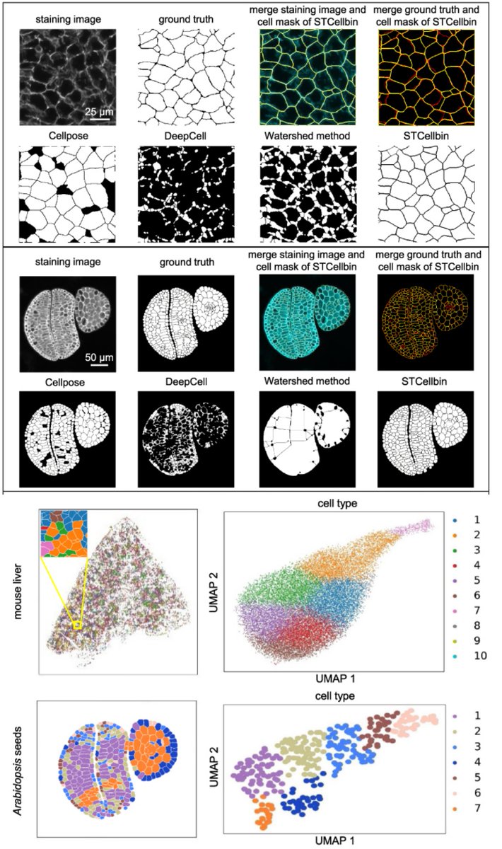

✨Our latest blog tests 8 popular community-developed tools using Stereo-seq data. Discover which algorithms excel across diverse staining types and explore segmentation-free methods for special cases! Dive in: bit.ly/3Vnn2D7 #CellSegmentation #Spatialbiology #STOmics


Working on cell segmentation in colon tissue @MikeKattahMD @iuliarusu9 @JaredBain909 #cellsegmentation #spatialbiology #colon #colorful #ucsf


We are very impressed with the ease of creation and quality of this open source @DeepCell_Inc #CellSegmentation mask made in Steinbock in the @BodenmillerLab. Our #ImagingMassCytometry data analysis is much improved!

Welcome to the 6th day of 12 Days of NanoString! Interested in leaping to single-cell spatial in these applications? nanostring.com/events/spatial… #SpatialData #CellSegmentation #SpatialTranscriptomics #SingleCellImaging


DON'T MISS Dr. McArdle's @pmlsummit talk this Thurs 9/14 featuring Orion data utilizing @QuPath for #imagevisualization, #cellsegmentation via #deeplearningmodels, #cellphenotyping, and #spatialanalysis. For free registration: rarecyte.com/contact-us/ #spatialbiology


Ko Sugawara shows how training with sparse annotations can produce practical models for cell image segmentation. Strategic selection of annotated cells boosts performance! @ELEPHANT_track @NEUBIAS_ Symposium 🧪🔍 #DeepLearning #BioimageAnalysis #CellSegmentation

Dr. McArdle's @pmlsummit talk on 9/14 will feature Orion data being used with @QuPath for #imagevisualization, #cellsegmentation via #deeplearningmodels, #cellphenotyping, and #spatialanalysis. For free conference registration: rarecyte.com/contact-us/ #spatialbiology


RAMCES, a computational method by @monica_dayao et al., improves #cellsegmentation in imaging-based #spatialproteomics by facilitating the selection of optimal membrane protein markers @CMUPittCompBio @CMUCompBio go.nature.com/37TVoZg

The product of the study ""Real-Time Three-Dimensional #cellsegmentation in Large-Scale Microscopy Data of Developing #embryos"" (available at sciencedirect.com/science/articl…) can be observed throught #MorphoNet. #imagevisualization #CellBiology 🔬

Did you know? The @EnableMedicine platform allows you to visualize and interpret your #cellsegmentation results, with no coding involved! Upload your cell segmentation masks directly onto the @EnableMedicine platform, and view the masks and data on the Visualizer, with easy…
✨Our latest blog tests 8 popular community-developed tools using Stereo-seq data. Discover which algorithms excel across diverse staining types and explore segmentation-free methods for special cases! Dive in: bit.ly/3Vnn2D7 #CellSegmentation #Spatialbiology #STOmics
Customize your #CellSegmentation with the new #Cellpose2 Plugin for the #Vizgen Post-Processing Tool! Visit our website to learn how you can improve segmentation results for challenging tissues with the plugin for #VPT: hubs.ly/Q02mN7ZV0 #MERSCOPE #MERFISH #SpatialOmics
Customize your #CellSegmentation with the new #Cellpose2 Plugin for the #Vizgen Post-Processing Tool! Visit our website to learn how you can improve segmentation results for challenging tissues with the plugin for #VPT: hubs.ly/Q02k2S000 #MERSCOPE #MERFISH #Spatialomics

#cellsegmentation and classification are critical tasks in spatial #omics data analysis. Unlike the typical two-stage approach of segmentation followed by classification, #CelloType implements multitask learning strategy that integrates the two, enhancing the performance of both.
🎯 Precise #CellSegmentation is challenging. @vizgen_inc uses cell boundary stains and robust algorithms for accuracy. See these #DataImages of FFPE mouse breast tumor by researchers at @NeuroAlc, showcasing cell boundary staining and segmentation revealing natural cell shapes.
🎯 Precise #CellSegmentation is challenging. @vizgen_inc uses cell boundary stains and robust algorithms for accuracy. See these #DataImages of FFPE mouse breast tumor by researchers at @NeuroAlc, showcasing cell boundary staining and segmentation revealing natural cell shapes.
💻Webinar Alert for #SpatialTranscriptomics Customers in Japan! Developments in Spatial Genomics Analysis and Advanced Cell Segmentation 📅 Wednesday, April 24, 2024 ⏰15:00 - 15:45 JST 🎙️Wenbin Gu, Primetech Co. 🔗 hubs.ly/Q02tBwP_0 #CellSegmentation #MERSCOPE

💻Webinar Alert for #SpatialTranscriptomics Customers in Japan! Developments in Spatial Genomics Analysis and Advanced Cell Segmentation 📅 Wednesday, April 24, 2024 ⏰15:00 - 15:45 JST 🎙️Wenbin Gu, Primetech Co. 🔗 hubs.ly/Q02sQSrr0 #CellSegmentation #MERSCOPE
STCellbin Use Nuclei+Cell Membrane/Wall stains to improve #CellSegmentation & single-cell-resolved #SpatialTranscriptomics Tailored for #StereoSeq, but likely implementable for other ST methods Susanne Brix & Xun Xu labs @GigaByteJournal 2024 gigabytejournal.com/articles/110

🔬 Reducing annotation costs in #cellsegmentation📉 Study shows how a small, annotated dataset can train a model to upgrade low-quality annotations, improving segmentation accuracy and efficiency. 💡mdpi.com/2313-433X/10/7… #MDPIJimaging #DeepLearning #ComputerVision

Customize your #CellSegmentation with the new #Cellpose2 Plugin for the Vizgen Post-Processing Tool! Visit our website to learn more about #VPT and the Cellpose2 cell segmentation plugin: hubs.ly/Q02m2Xf80 #Vizgen #MERSCOPE #MERFISH #Spatialomics


🔬 Reducing annotation costs in #cellsegmentation📉 Study shows how a small, annotated dataset can train a model to upgrade low-quality annotations, improving segmentation accuracy and efficiency. 💡mdpi.com/2313-433X/10/7… #MDPIJimaging #DeepLearning #ComputerVision


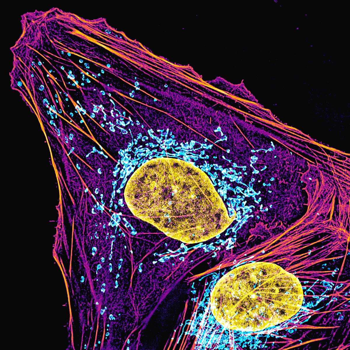
A cell photographed through a microscope. DNA (yellow), mitochondria (blue) and actin filaments (purple) are shown. Gamma correction on the actin filament channel was used to simultaneously display both the bright stress fibers and the dim cortex. #CellBiology #SciArt #Biology


LIVE LONGER YOUNGER - The Miracle Within. This is the most detailed image ever captured of a human cell, and it is absolutely breathtaking. Every color, every structure, every microscopic detail represents life in motion. Millions of signals. PM me for info.


These are the most detailed images of a human cell to date, obtained by radiography, nuclear magnetic resonance and cryoelectron microscopy.


In October '21, researchers from @Harvard, @BostonChildrens, & @uni_copenhagen published a paper in @NatureBiotech describing a new method for #CellSegmentation called #Baysor. Read & learn how they evaluated it with #MERFISH! hubs.ly/Q013pM8Q0 #ThrowbackThursday
#Baysor #CellSegmentation for 2D 3D image-based #SpatialTranscriptomics Based on Transcript composition #MarkovRandomField -/+Stain image #PriorSegmentationConfidence Work with #MERFISH #smFISH #STARmap #InSituSequencing ⏫Detection of #EndothelialCell #MuralCell (Fig 5/6)…
STCellbin Use Nuclei+Cell Membrane/Wall stains to improve #CellSegmentation & single-cell-resolved #SpatialTranscriptomics Tailored for #StereoSeq, but likely implementable for other ST methods Susanne Brix & Xun Xu labs @GigaByteJournal 2024 gigabytejournal.com/articles/110
💻Webinar Alert for #SpatialTranscriptomics Customers in Japan! Developments in Spatial Genomics Analysis and Advanced Cell Segmentation 📅 Wednesday, April 24, 2024 ⏰15:00 - 15:45 JST 🎙️Wenbin Gu, Primetech Co. 🔗 hubs.ly/Q02sQSrr0 #CellSegmentation #MERSCOPE
A cell undergoing cell division photographed through a microscope. Actin filaments (red) and myosin II filaments (cyan) are shown. #CellBiology

✨Our latest blog tests 8 popular community-developed tools using Stereo-seq data. Discover which algorithms excel across diverse staining types and explore segmentation-free methods for special cases! Dive in: bit.ly/3Vnn2D7 #CellSegmentation #Spatialbiology #STOmics

It's not AI. It's not a painting. It is the most detailed image of a human cell to date, obtained by radiography, nuclear magnetic resonance and cryoelectron microscopy. _____ Original post: linkedin.com/posts/laurent-…


AI generated histology derived from OCT Fascinating images #TCT2025 Learning pathology through OCT! @ziadalinyc @GreggWStone @crfheart @JEscaned @PCRonline

A dividing cancer cell photographed with a microscope. Actin filaments, microtubules and DNA are shown. #CellBiology #microscopy #SciArt

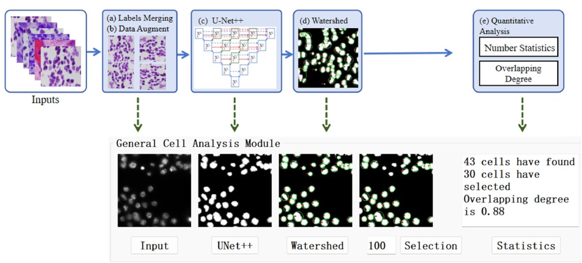
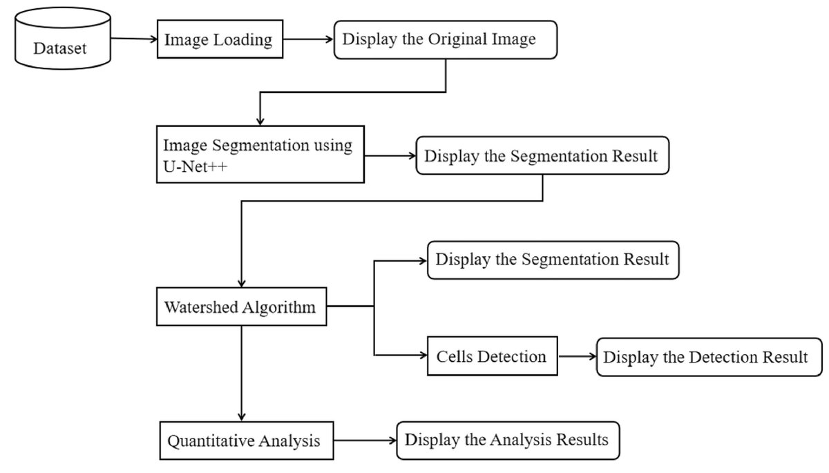
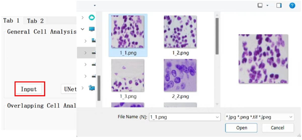
Keywords Image Segmentation, Deep Learning, Image Analysis, U-Net++, Watershed, GUI
Get a closer look at the superior cell segmentation and transcript information you can obtain from CosMx SMI data during our upcoming Spatial Informatics 101 Webinar on July 11. Register to attend: bit.ly/3pxrlQ4 #SpatialInformatics #CellSegmentation

Cell shading style!!!!! I think that's what it's called ??


Customize your #CellSegmentation with the new #Cellpose2 Plugin for the Vizgen Post-Processing Tool! Visit our website to learn more about #VPT and the Cellpose2 cell segmentation plugin: hubs.ly/Q02m2Xf80 #Vizgen #MERSCOPE #MERFISH #Spatialomics
Something went wrong.
Something went wrong.
United States Trends
- 1. Clippers 14K posts
- 2. Harden 10.2K posts
- 3. Huda 8,969 posts
- 4. #DWTS 50.1K posts
- 5. Ty Lue 1,505 posts
- 6. Kawhi 3,649 posts
- 7. Giannis 30K posts
- 8. Whitney 15.6K posts
- 9. #RHOSLC 7,754 posts
- 10. #APEC2025 77.7K posts
- 11. #RMxAPEC 191K posts
- 12. #DearOlandria 1,147 posts
- 13. Askarov N/A
- 14. South Korea 72.8K posts
- 15. Markstrom 1,723 posts
- 16. Caden 8,431 posts
- 17. Wayne 57.7K posts
- 18. Chris Paul 1,545 posts
- 19. Zubac 1,699 posts
- 20. Joon 13.3K posts