#lightsheet search results

🚨Big news! Five years in the making, our Zebrahub paper is now published in #Cell 🎉. We’ve built a timecourse atlas of zebrafish embryonic development, combining #scRNAseq data and #lightsheet live imaging, and delved deep into the dynamics of key progenitors.…
We present #Zebrahub: a timecourse atlas of zebrafish embryonic development, combining #scRNAseq time-course data with #lightsheet live imaging. Explore our seq. and imaging datasets interactively at biorxiv.org/content/10.110… zebrahub.org 1/n

How many #lightsheet systems are out there that can be set up within 3 minutes (including booting up the laptop), fit into your hand luggage, and still are fun to operate? Thanks for the fun time today at the open hardware workshop @Chagas_AM et al.! :) #imswitch @xaviercasasm

#zebrafish larvae imaged on home-built #lightsheet microscope assembled by a team of remarkable students Shawniya, Josh @joshsselfe, Stephan and Syed @SyedAdnanUddi11 with the guidance of incredible researchers @HagarLavian and @PortuguesLab at @CSHL @cshlcourses #neuroscience…


🚨🔬Ready to push the limits of advanced bioimaging? Our team has two job openings at Chan Zuckerberg Biohub in #SanFrancisco! czbiohub.org/royer/ #lightsheet, #microscopy, #optics, #zebrafish and more. The first position is for a recent Ph.D. graduate who will design…

My colleagues put together an ebook on tissue clearing (for #lightsheet microscopy). If you are like me and have only experience with live imaging and are a bit confused with the different acronyms flying around, I highly recommend you take a look! I counted 37 methods!


Excited to share work by @SarahAEYoung featuring #cover @ScienceAdvances, showing with #lightsheet and #microCT cancer cells homing to all bone sites, yet metastasis initiating in high remodeling sites. @MpiciPotsdam @biogipuzkoa @Ikerbasque @dfg_public 👉bit.ly/3UQXvTW


Next week we will be attending the @BaCell3D on May 8-9 in #Basel. Come and talk to us to know more about our latest LS2 Live system and how we image organoids for extended period of time using #lightsheet. Video credit @FranziskaMoos, @priscaliberali lab.
The latest work from @OatesLab @EPFL_en shows how cells are instructed from the segmentation clock to form somite boundaries in #zebrafish embryo. biorxiv.org/content/10.110… Check out this wonderful movie acquired on our #lightsheet by lead author @Olivier_Venzin.

Hey nerds, meet Astyanax Mexicanus. Surface dweller has eyes, bottom dweller not. Critter's brain cleared then imaged with a #lightsheet #microscope from @3i_inc. @JohannaKowalko Lab @LehighU. Sample courtesy of @ReneeMapa. Keep your eyes on Renee. Happy #FluorescenceFriday

The beautiful work from @diane_pelzer in @maitrejl Maître's lab in now published in @emboJournal. Our LS1 Live #lightsheet was instrumental to investigate the mechanism behind cell fragmentation in pre-implantation #embryos. embopress.org/doi/full/10.15…
It took 4+ years to build this 3D map of the developing human head, the most complex part of our body sciencedirect.com/science/articl… Tissue clearing #Lightsheet #VirtualReality #3D models & More! Congrats to Raphael Blain, E. Shotar G. Couly @Yorick_Gitton et al. video summary 🔊

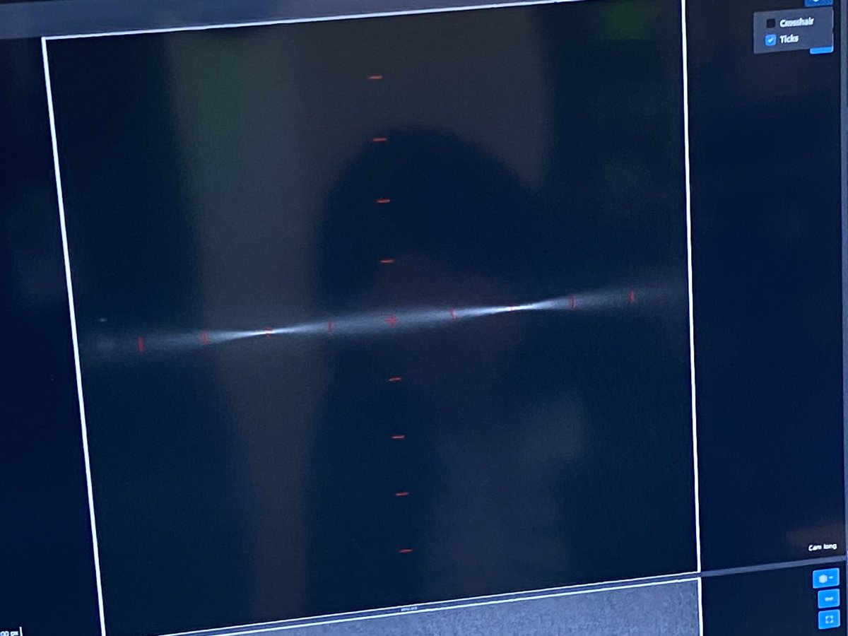
After a long #roadtrip through the USA, the last conference on computational optics @HHMIJanelia is almost over. Just a quick last measurement before going to bed in ordr to check the deconv from @optrickster's lab using the @openuc2 #lightsheet using #ImSwitch. Works ☑️

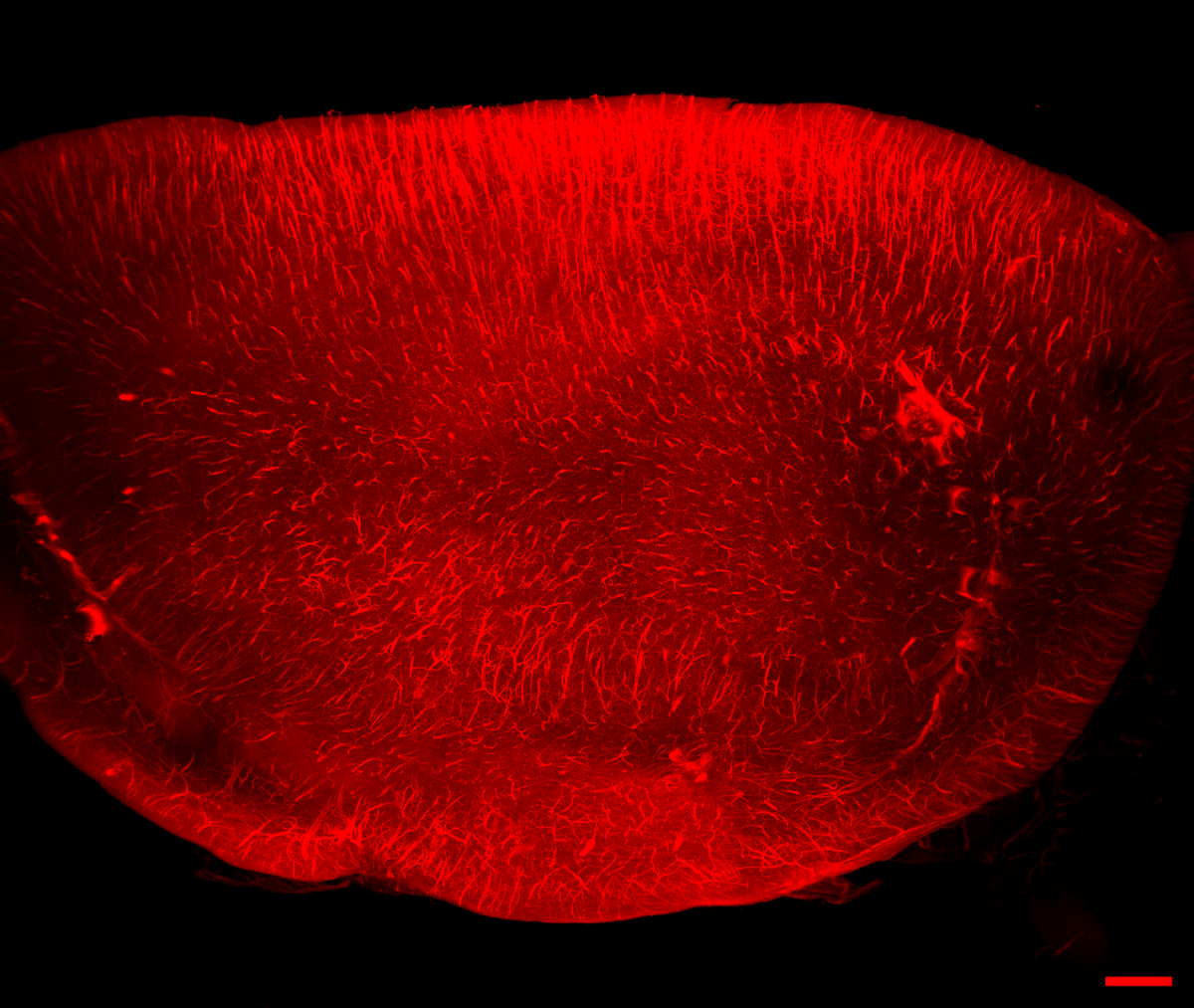
First #LightSheet from my lab. Retinal organoid with Müller glia (cyan), Retinal Ganglion Cells (red). Without #imaris, just #ImageJ, #Fiji, and #3Dscript. With help of @Peta_Machackova from @Ceitec_CellimCF done using #EZClear method (@wythelab) by #PhDstudent Canan Celiker

Last week I had the privilege of visiting the amazing facilities of @mpicbg, including a sneak peek of their OpenSPIM. Many thanks to my excellent guides @PavelTomancak and Jan Peychl! #lightsheet #microscopy


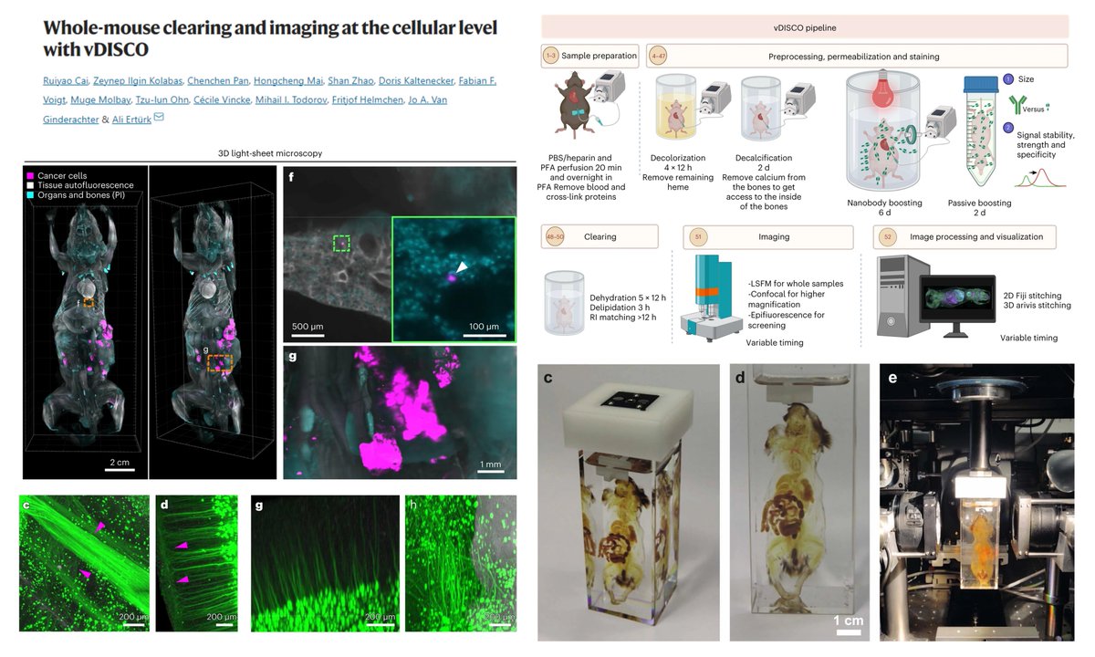
vDISCO Optically clear the whole mouse & image at single-cell level #LightSheet #mesoSPIM A great list of vDISCO-compatible Nanobodies (Table 1) +Decolorization RBC +Decalcification Bone Dr. Ali Ertürk lab @erturklab @NatureProtocols 2023 @MarikaRuiyao nature.com/articles/s4159…

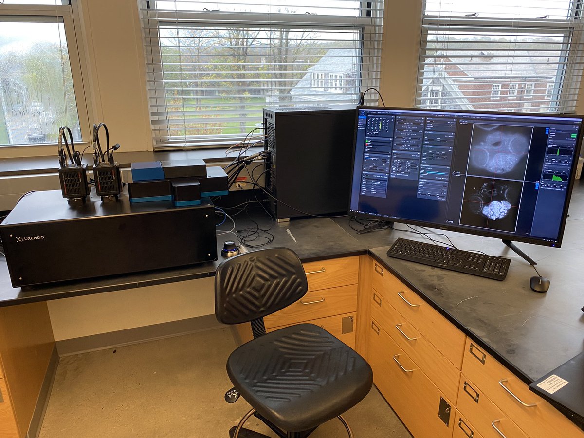
We're happy to be part of #tim2023 organized by @GerBI_GMB with one of our T-SPIM Flamingo #lightsheet microscopes. Very interesting discussions and lots of input!

Proud to be part of this transformative work from the Tomer Lab & @mbfbioscience.bsky.social 🔬 HySIL & SCOPE technologies power this new SLICE light-sheet microscope. 🚀 Submicron resolution, multi-immersion, and affordability. biorxiv.org/content/10.110… #Microscopy #LightSheet
Big data in microscopy shouldn't be a bottleneck. I started Arnas Technologies arnastech.com to solve exactly that. We build custom #AI workflows to automate analysis for #lightsheet, #microscopy, and #OMICS data. #BioTech #DataScience #ImageAnalysis #BioImageAnalysis


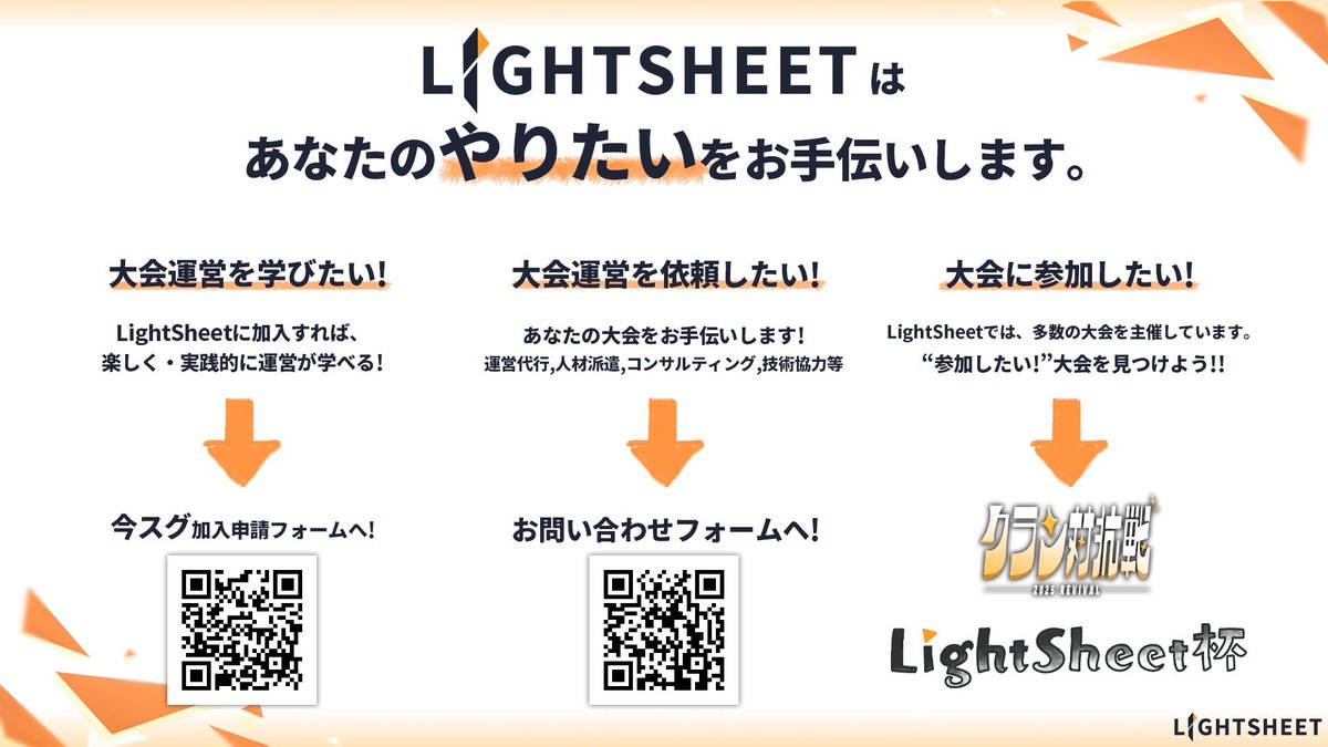
第13回Light sheet杯 結果準優勝でした! ハピハロでめっちゃやらかしたしなんで決勝相手身内なん? めっちゃ悔しいので優勝するまで大会で続けたいと思います #Lightsheet #Lightsheet杯 #プロセカ

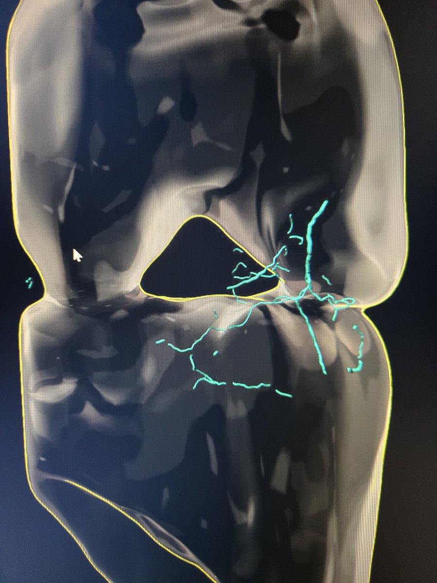
Glomeruli, the filtering parts of nephrons, are visible in cyan in this mouse kidney section imaged with #DALISPIM. Our new open-top #lightsheet microscope's unique design allowed us to image this 1-mm thick sample directly in a 12-well plate. #nephrology
The days are getting darker, so we're celebrating what we can do with light 💡 Come hang out with us at the New England Society for Microscopy's Fall Symposium @BrandeisU this Thursday & Friday. We'll be ready to talk all things #lightsheet microscopy!

Want to see high-resolution #lightsheet datasets from a diverse collection of species and tissue types in one place? 🐠🐁🦎🔬 Visit our video gallery to see all of our past data drops, and bookmark it to keep up with new ones: hubs.ly/Q03PH7VF0


#HighlyAccessedPaper "Three-Dimensional Histological Characterization of the Placental Vasculature Using Light Sheet Microscopy" by Lennart Freise, et al. Cited 3 times | Viewed 3797 times. Read now ➡️ brnw.ch/21wWPQ6 #LightSheet #Placenta #Preeclampsia #Vascular

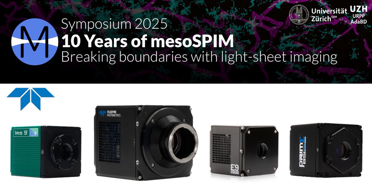
We’re heading to Zürich for the #mesoSPIM 10-Year Symposium, Oct 13–15th! Join us as we celebrate a decade of open-source #lightsheet #microscopy and explore the future of high-speed, high-resolution imaging. Come chat with us about the latest in #scientificcameras!

「大会運営って、何から始めれば…?」 LightSheetなら、運営のノウハウをゼロから学べる研修(LightSheet杯)や、スキルアップのための講習会が充実しています! 未経験からでも、安心して"創る側"に挑戦できます。 少しでも興味を持ったら、ぜひお話だけでも聞きに来てください! #LightSheet

#Mousebrain with neurons from the eyes. This cleared 870 GB tiled z-stack from a @zeiss_micro #LightSheet Z.1 was deconvolved, stitched, and vignetting corrected w\ Huygens. c/o: Dominic Fillion, Montreal Clinical Research Institute Webinar September 16: svi.nl/webinarinvitat…

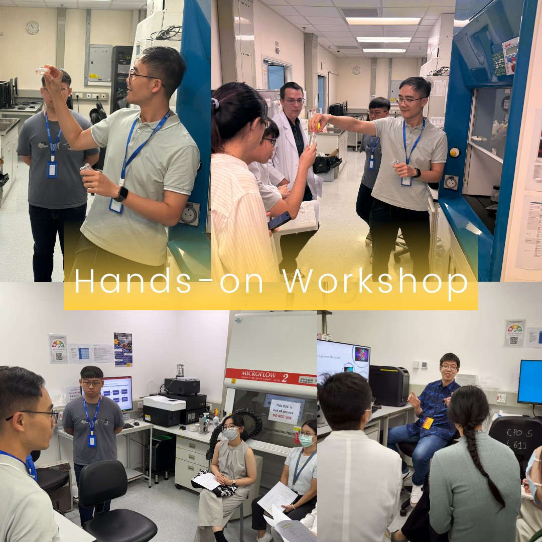
5-day Workshop for Tissue Clearing for Light-sheet Microscopy🧪✨ Big thanks to Prof. Lai and Dr. Zhao for sharing their knowledge! 👉Swipe for workshop highlights👉 #LightSheet #3DImaging #TissueClearing #LiToneXLLightsheetMicroscope #LightsheetMicroscopy #ImagingCytometry


Check out this interview by @zeiss_micro with @anvargasr, our initiative's coordinator, on building a South American #lightsheet community and our experience so far. Transforming South American Research: Core Imaging Facilities and Light-Sheet Microscopy zeiss.com/microscopy/en/…

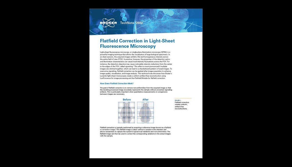
🔬 Flatfield Correction in Light-Sheet Fluorescence Microscopy In this technical note: 🔹 The purpose of flatfield correction 🔹 Steps to take for applying flatfield correction 🔹 Application example 🔗 buff.ly/3AM50Un #lsfm #spim #lightsheet #microscopy
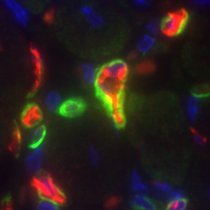
#Photomanipulation with #LightSheet? Yes, indeed. Data show #zebrafish microglia and macrophage dynamics after small injury induced by photomanipulation (right), compared to the control fish (left). Thank you Gordon Wang for the samples. @bruker Luxendo TruLive3D Imager


#zebrafish larvae imaged on home-built #lightsheet microscope assembled by a team of remarkable students Shawniya, Josh @joshsselfe, Stephan and Syed @SyedAdnanUddi11 with the guidance of incredible researchers @HagarLavian and @PortuguesLab at @CSHL @cshlcourses #neuroscience…
My colleagues put together an ebook on tissue clearing (for #lightsheet microscopy). If you are like me and have only experience with live imaging and are a bit confused with the different acronyms flying around, I highly recommend you take a look! I counted 37 methods!

Last cell phone pic, I promise! Full rendering is coming soon. #tissueclearing #lightsheet #EZClear #OiVM

Our #MegaSPIM microscope is installed and ready to go for the @MicRoNHMS demo! Excited to work with Drs. Anekal, @LlopisMontero, & Wells to generate some awesome #lightsheet data 🔬

Excited to share work by @SarahAEYoung featuring #cover @ScienceAdvances, showing with #lightsheet and #microCT cancer cells homing to all bone sites, yet metastasis initiating in high remodeling sites. @MpiciPotsdam @biogipuzkoa @Ikerbasque @dfg_public 👉bit.ly/3UQXvTW
After a long #roadtrip through the USA, the last conference on computational optics @HHMIJanelia is almost over. Just a quick last measurement before going to bed in ordr to check the deconv from @optrickster's lab using the @openuc2 #lightsheet using #ImSwitch. Works ☑️
We're happy to be part of #tim2023 organized by @GerBI_GMB with one of our T-SPIM Flamingo #lightsheet microscopes. Very interesting discussions and lots of input!

Welcome to our new colleague Sona Legartova! She has a lot of experiences with microscopy and will be a great asset to our users. Already starting with #lightsheet microscopy.

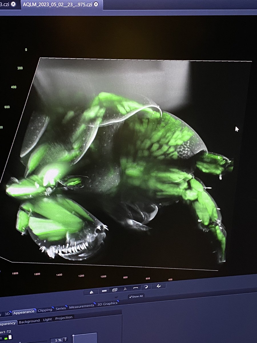
Guys this is a #transgenic #crustacean 😍🤯 Imaged on @zeiss_micro #lightsheet 7 #microscope #aqlm2023 @AQLMMBL @MBLScience #microscopy
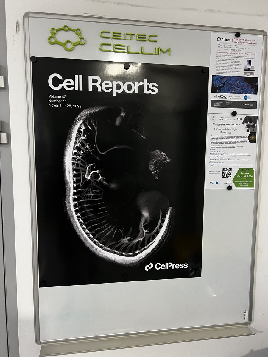
Our new old school analog notice board. With amazing cover picture from our #lightsheet @zeiss_micro 🔬. Fantastic work of @AlcaSalas @KrivanekLab.
Hey nerds, meet Astyanax Mexicanus. Surface dweller has eyes, bottom dweller not. Critter's brain cleared then imaged with a #lightsheet #microscope from @3i_inc. @JohannaKowalko Lab @LehighU. Sample courtesy of @ReneeMapa. Keep your eyes on Renee. Happy #FluorescenceFriday
Last week I had the privilege of visiting the amazing facilities of @mpicbg, including a sneak peek of their OpenSPIM. Many thanks to my excellent guides @PavelTomancak and Jan Peychl! #lightsheet #microscopy
We’re heading to Zürich for the #mesoSPIM 10-Year Symposium, Oct 13–15th! Join us as we celebrate a decade of open-source #lightsheet #microscopy and explore the future of high-speed, high-resolution imaging. Come chat with us about the latest in #scientificcameras!
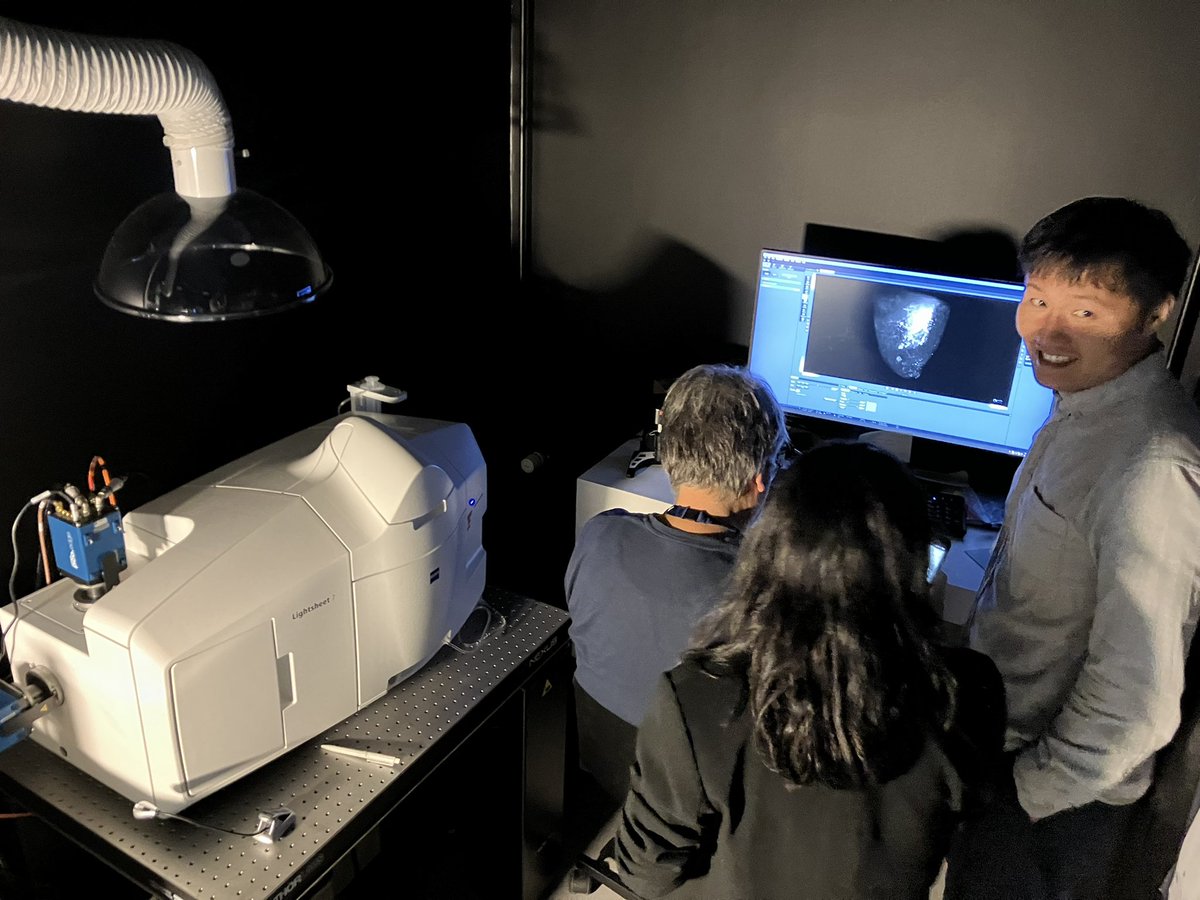
My summer break is over but the #lightsheet course @AarhusUni_int @DBioimaging is just as much fun by the sea :-) We brought an #Acquifer #HIVE for #imageprocessing of #largedata from @zeiss_micro #Lightsheet7 by 20 people in parallel — @luxendo @BrukerFM @zeiss_arivis


#expansion microscopy is fantastic tool. We can provide support through all procedure - sample preparation, image acquisition up to image processing and analysis. Here @Peta_Kucerova and @veronikapospis6 during #lightsheet 🔬 imaging.


Something went wrong.
Something went wrong.
United States Trends
- 1. Everton 127K posts
- 2. GeForce Season 3,079 posts
- 3. Comey 160K posts
- 4. Amorim 51.7K posts
- 5. Seton Hall 2,026 posts
- 6. Manchester United 76.1K posts
- 7. Mark Kelly 106K posts
- 8. Pickford 9,638 posts
- 9. #MUNEVE 14.9K posts
- 10. #MUFC 22.8K posts
- 11. Opus 4.5 7,415 posts
- 12. Dorgu 19.2K posts
- 13. UCMJ 15.8K posts
- 14. Zirkzee 22K posts
- 15. Hegseth 39.2K posts
- 16. Man U 33.5K posts
- 17. Amad 12.2K posts
- 18. Gueye 29.5K posts
- 19. Keane 18.1K posts
- 20. Will Wade N/A